How to Draw and Label a Human Heart Diagram, Step by Step
The human heart diagram is one of the most asked diagrams in Class 10 Biology (Life Processes). It shows the four chambers, the big blood vessels and the valves that keep blood moving one way. Below it is broken into small steps, so you build it up part by part instead of copying the whole thing at once.
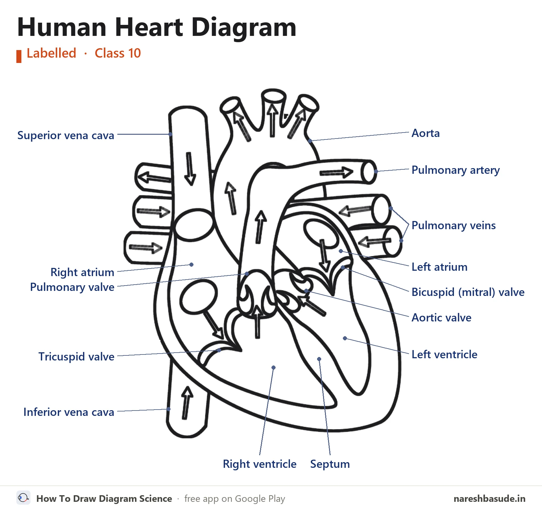
The finished human heart diagram with all 14 parts labelled. The steps below build it up one part at a time.
What the human heart does
The heart is a muscular pump with four chambers: two atria on top and two ventricles below. The right side takes in deoxygenated blood from the body and sends it to the lungs. The left side takes in oxygenated blood from the lungs and pumps it out to the whole body through the aorta.
Draw it in 9 steps
Use a sharp pencil and draw lightly at first. In every picture, red is what to draw in that step and grey is what you have already drawn.
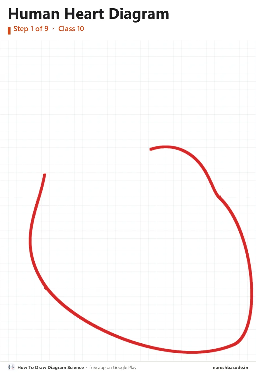
The heart's body
Draw the big body of the heart. Start at the top left, sweep down and round the bottom, then go up the right side. Keep it smooth, like a tilted U. The lowest point is a little to the right.
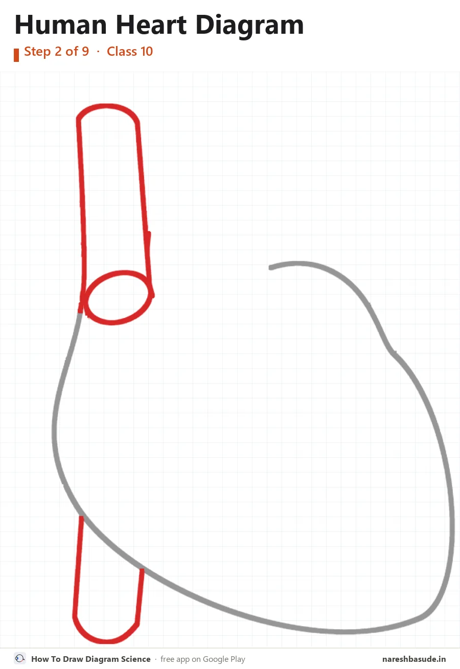
Vena cava
On the left, draw a tall tube coming down from the top, with an oval at its lower end. Then draw a short tube coming out at the bottom left.
Parts drawn: Superior vena cava (top tube), Inferior vena cava (bottom tube)
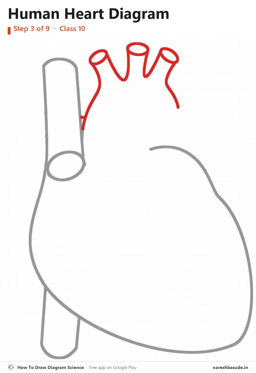
Aorta
Draw the aorta on top: a curved arch with three short branches pointing up. Give each branch an oval opening at its top.
Parts drawn: Aorta
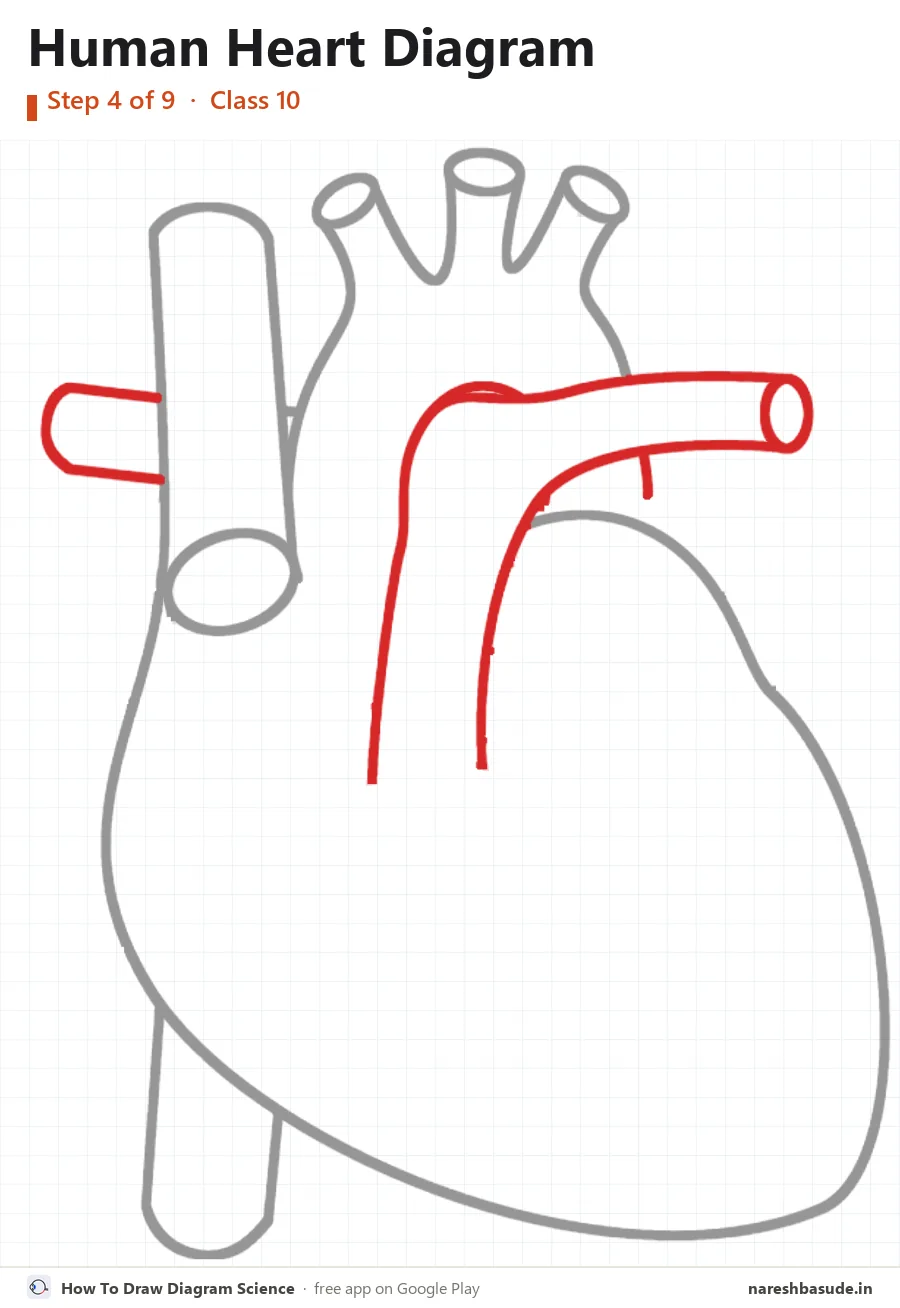
Pulmonary artery
From the middle, draw a thick tube going up and bending to the right, with an oval at its end. Then draw its other branch: a short tube going out to the left, behind the vena cava.
Parts drawn: Pulmonary artery
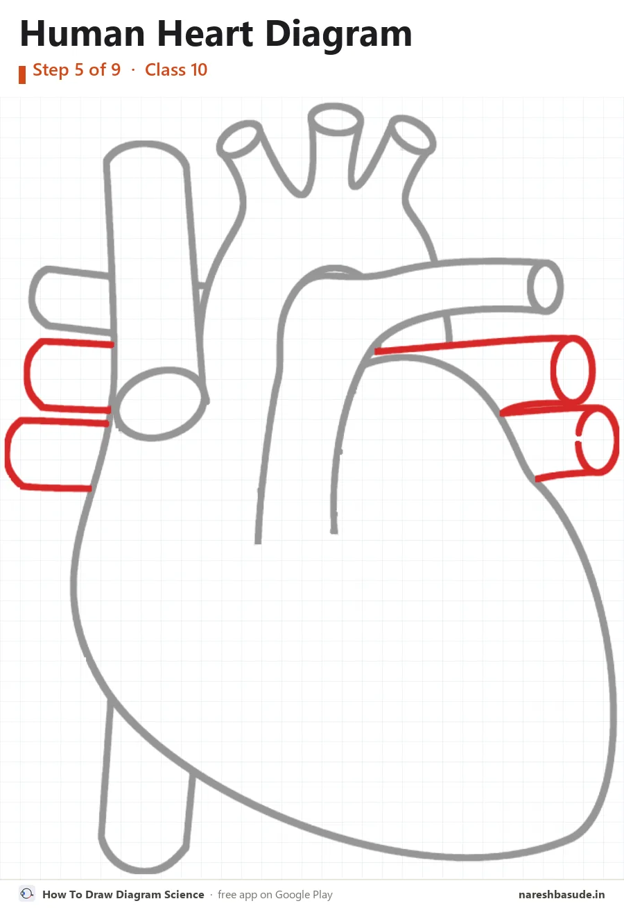
Pulmonary veins
Draw four short tubes: two on the right with ovals at their ends, and two on the left behind the vena cava. They bring blood back from the lungs.
Parts drawn: Pulmonary veins
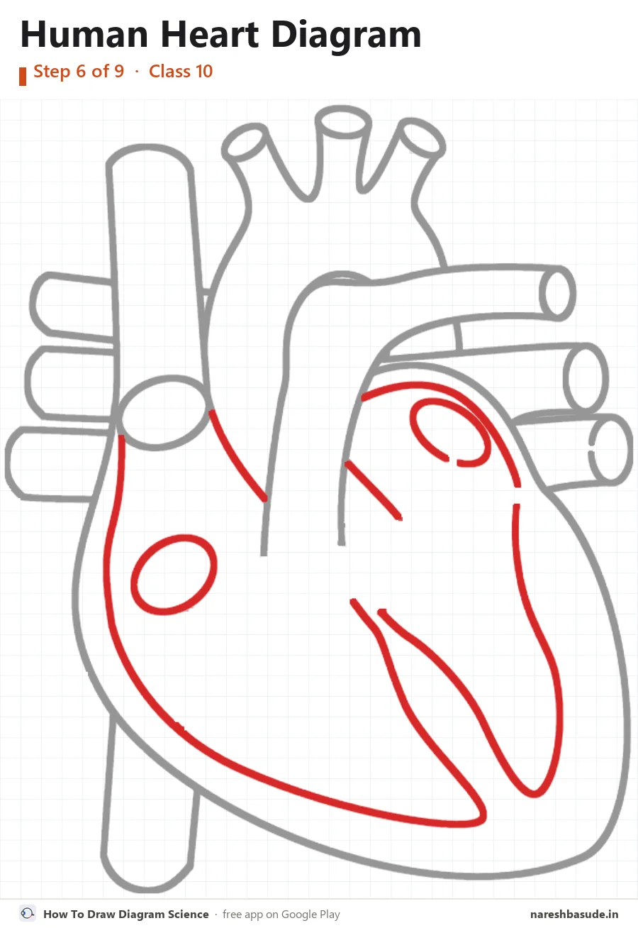
Inner walls
Now the inside. Draw the wall lines that make the four rooms: two atria on top and two ventricles below, with the septum wall down the middle. Add the two round openings.
Parts drawn: Right atrium, Left atrium, Right ventricle, Left ventricle, Septum
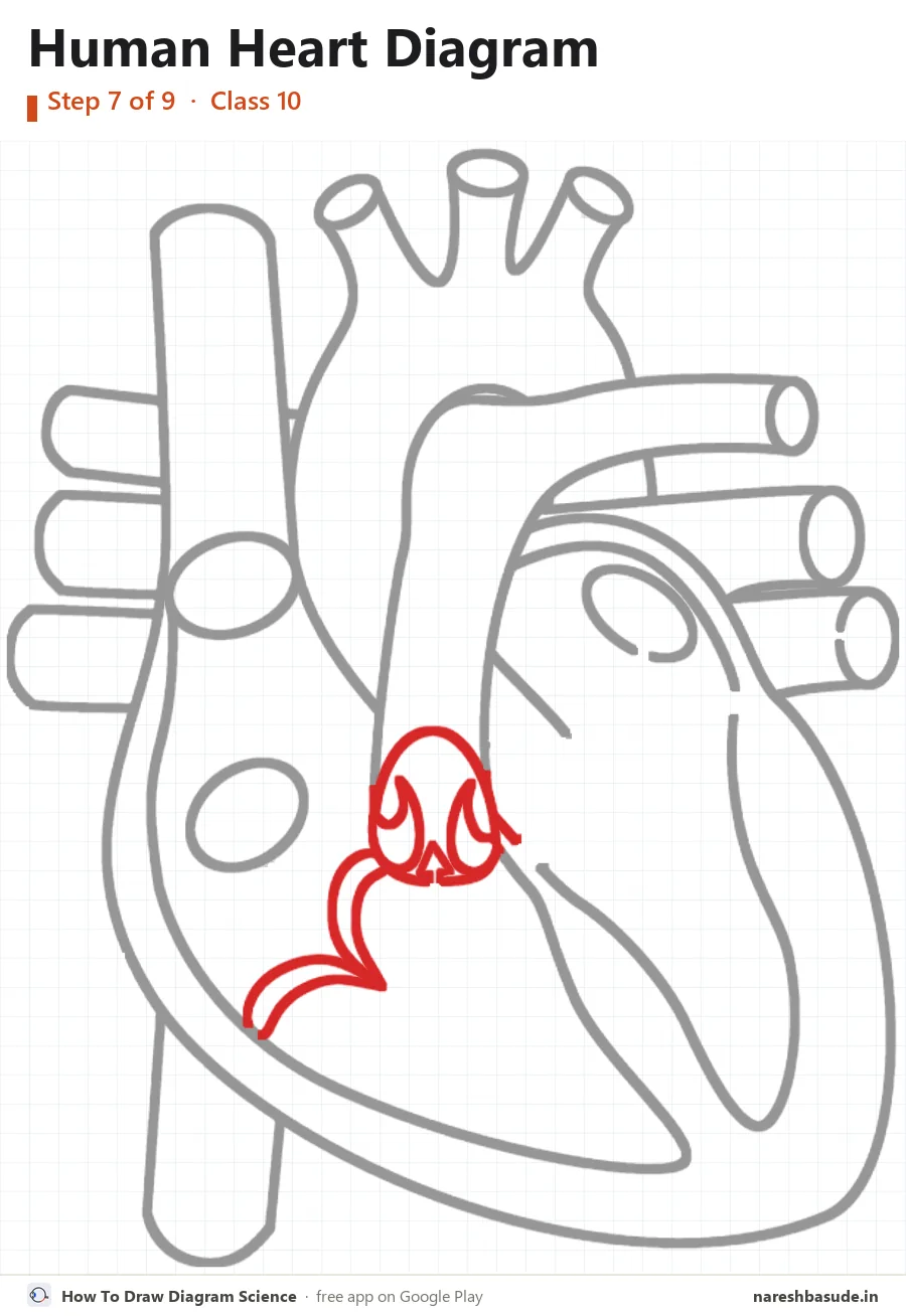
Valves on the right side
Draw the curled flap at the bottom left, then the round cup at the foot of the pulmonary artery with the small flaps inside it.
Parts drawn: Tricuspid valve, Pulmonary valve
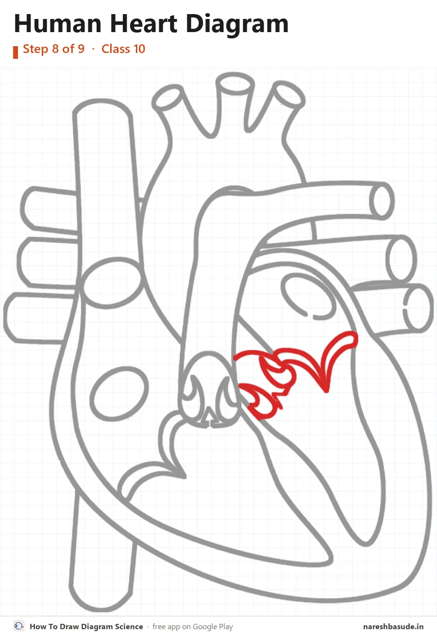
Valves on the left side
Draw the small curled flaps just right of the pulmonary valve, then the hooked flap further right.
Parts drawn: Aortic valve, Mitral (bicuspid) valve
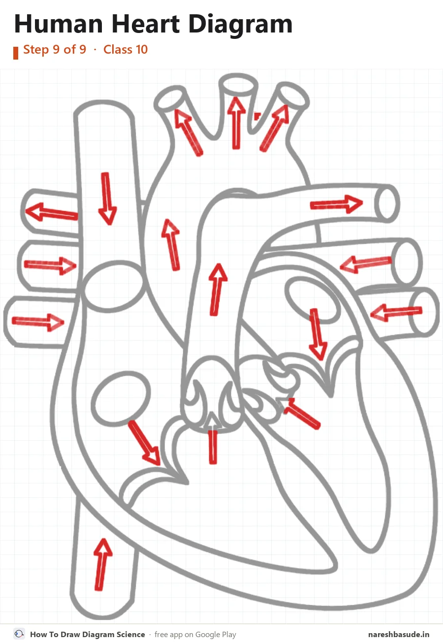
Blood-flow arrows
Add the arrows. In the veins they point into the heart; in the arteries they point out of the heart.

Parts to label (14) and what each one does
- Superior vena cava Brings deoxygenated blood from the head, neck and arms into the right atrium.
- Right atrium Upper right chamber. It receives deoxygenated blood from the body through the venae cavae.
- Pulmonary valve At the opening of the pulmonary artery. It stops blood flowing back into the right ventricle.
- Tricuspid valve The valve between the right atrium and the right ventricle. It stops blood flowing back into the atrium.
- Inferior vena cava Brings deoxygenated blood from the lower body into the right atrium.
- Aorta The largest artery. It carries oxygenated blood from the left ventricle to the whole body.
- Pulmonary artery Carries deoxygenated blood from the right ventricle to the lungs. It is the only artery that carries deoxygenated blood.
- Pulmonary veins Bring oxygenated blood from the lungs to the left atrium. They are the only veins that carry oxygenated blood.
- Left atrium Upper left chamber. It receives oxygenated blood from the lungs through the pulmonary veins.
- Bicuspid (mitral) valve The valve between the left atrium and the left ventricle. It stops blood flowing back into the atrium.
- Aortic valve At the opening of the aorta. It stops blood flowing back into the left ventricle.
- Left ventricle Lower left chamber with the thickest wall. It pumps oxygenated blood to the whole body through the aorta.
- Right ventricle Lower right chamber. It pumps deoxygenated blood to the lungs through the pulmonary artery.
- Septum The wall between the right and left sides of the heart, so oxygenated and deoxygenated blood do not mix.
Write each name on the side of the diagram, joined to its part with a straight line. Keep the lines from crossing.
Human Heart diagram without labelling (practice sheet)
The same diagram with the label lines but no names (an unlabelled diagram). Write each part's name on its line, then check your answers against the labelled diagram at the top.
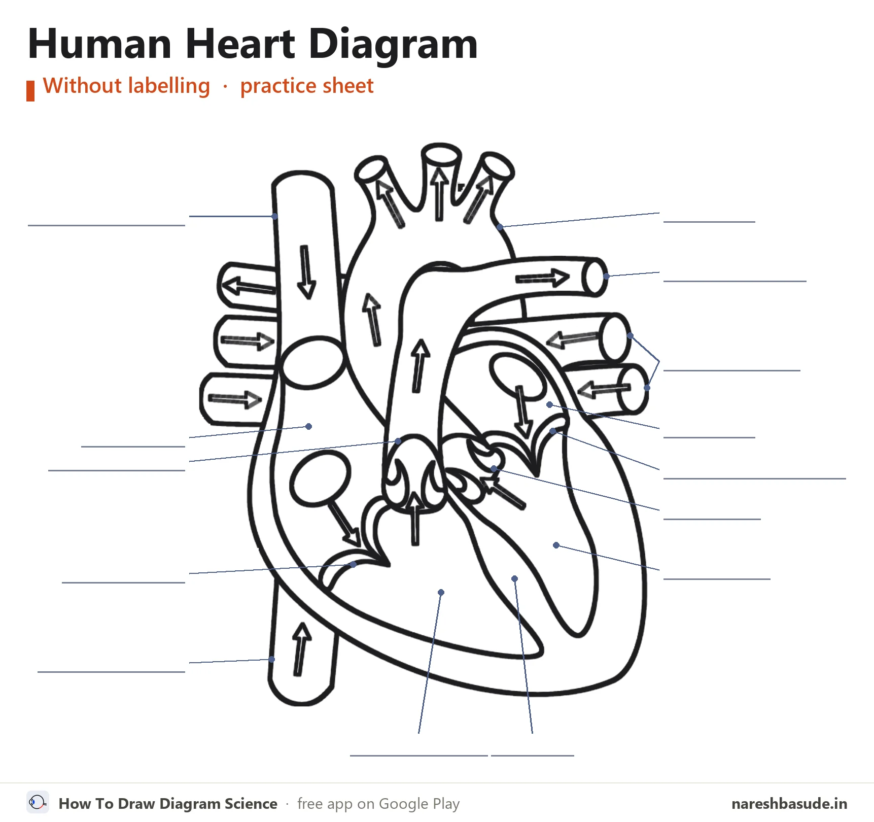
Download free PDF (A4, 3 pages)
Page 1: the labelled diagram. Page 2: this practice sheet with a word bank of all 14 parts. Page 3: all 9 drawing steps on one page.
Exam tips
- Draw the heart as if the person is facing you. That is why the heart's right side (right atrium, right ventricle) sits on your left.
- Blood vessels first, inner walls after. Once the outer tubes are in place, the four chambers and the septum are easy to fit inside.
- Arrows matter: in veins they point into the heart, in arteries they point out of it.
- Keep every label line straight and horizontal, with all names on the two sides. Lines that cross each other cost neatness marks.
Common mistakes
- Swapping the left and right sides of the heart.
- Mixing up the pulmonary artery (takes blood to the lungs) and the pulmonary veins (bring blood back from the lungs).
- Putting the bicuspid valve on the right. The tricuspid valve is on the right side; the bicuspid (mitral) valve is on the left.
- Forgetting the septum, the wall that separates the two ventricles.
Exam questions on the human heart diagram
QuestionDraw a neat diagram of the sectional view of the human heart and label: aorta, pulmonary artery, right atrium and left ventricle.
Draw the four-chambered heart as seen from the front. The aorta is the big arch at the top, coming out of the left ventricle. The pulmonary artery leaves the right ventricle just in front of it and goes to the lungs. The right atrium is the upper chamber on your left. The left ventricle is the thick-walled lower chamber on your right.
QuestionWhy is the wall of the left ventricle thicker than the wall of the right ventricle?
The left ventricle pumps blood to the whole body, which needs much more force than the right ventricle needs to send blood only to the lungs. So the left ventricle has a thicker muscular wall.
Questions students ask
How many chambers does the human heart have?
Four: two atria (the upper chambers) and two ventricles (the lower chambers).
Which blood vessel carries oxygenated blood from the heart to the body?
The aorta. It leaves the left ventricle and branches to carry blood all over the body.
Why is the right side of the heart drawn on the left?
Diagrams show the heart the way you would see it in a person standing in front of you, so their right side is on your left.
Practise it on your phone
These steps come straight from the free Android app How To Draw Diagram Science. In the app you can view the human heart with every part named, learn to draw it step by step on a numbered grid, take a quiz on the parts and label it yourself.


