How to Draw and Label a Human Digestive System Diagram, Step by Step
The human digestive system is a favourite in Class 10 Biology (Life Processes), and it is long, so a lot of students lose time on it. Broken into the small steps below, it is just a food pipe, a stomach, a few glands and two intestines drawn in order.
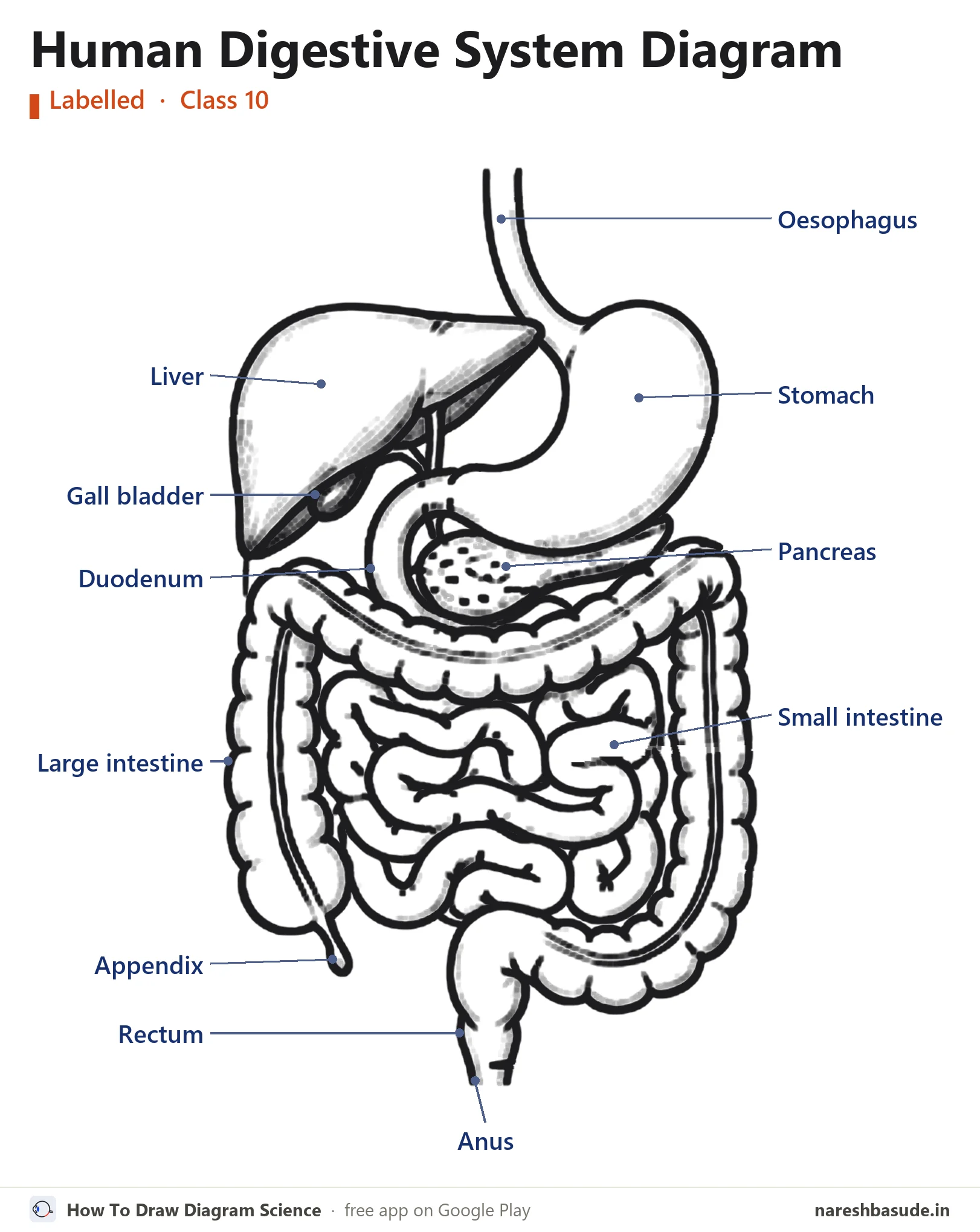
The finished human digestive system diagram with all 11 parts labelled. The steps below build it up one part at a time.
What the human digestive system does
Food passes from the mouth down the oesophagus to the stomach, then into the small intestine, where digestion is completed and food is absorbed. The large intestine absorbs water, and waste leaves through the rectum and anus. The liver makes bile, which is stored in the gall bladder; the pancreas adds pancreatic juice.
Draw it in 13 steps
Use a sharp pencil and draw lightly at first. In every picture, red is what to draw in that step and grey is what you have already drawn.
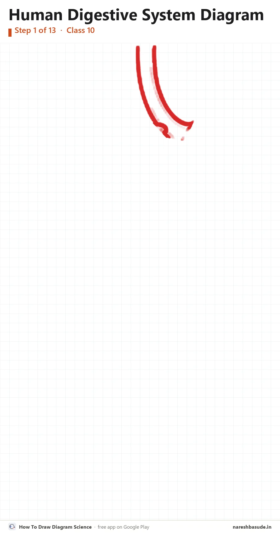
Oesophagus
Start at the top: draw the food pipe, a narrow tube coming down and bending to the right.
Parts drawn: Oesophagus
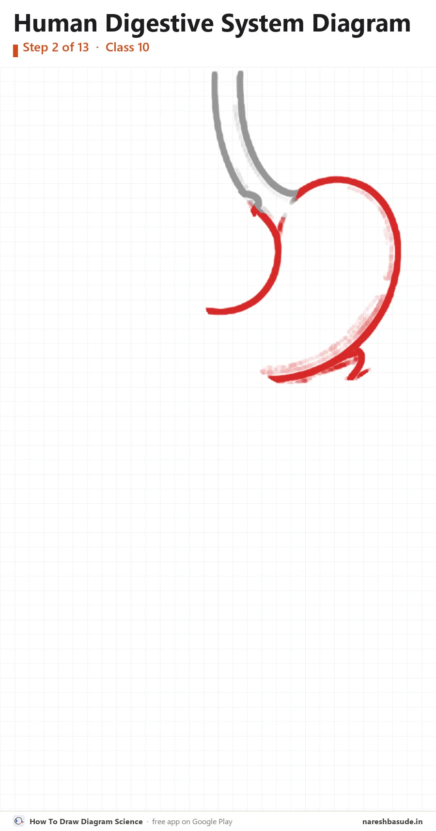
Stomach
At the end of the oesophagus draw the stomach: a large J-shaped bag on the right side of the picture.
Parts drawn: Stomach
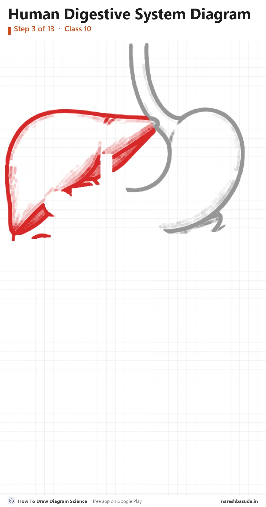
Liver
On the left draw the liver: the biggest organ, a wide wedge shape with a rounded top.
Parts drawn: Liver
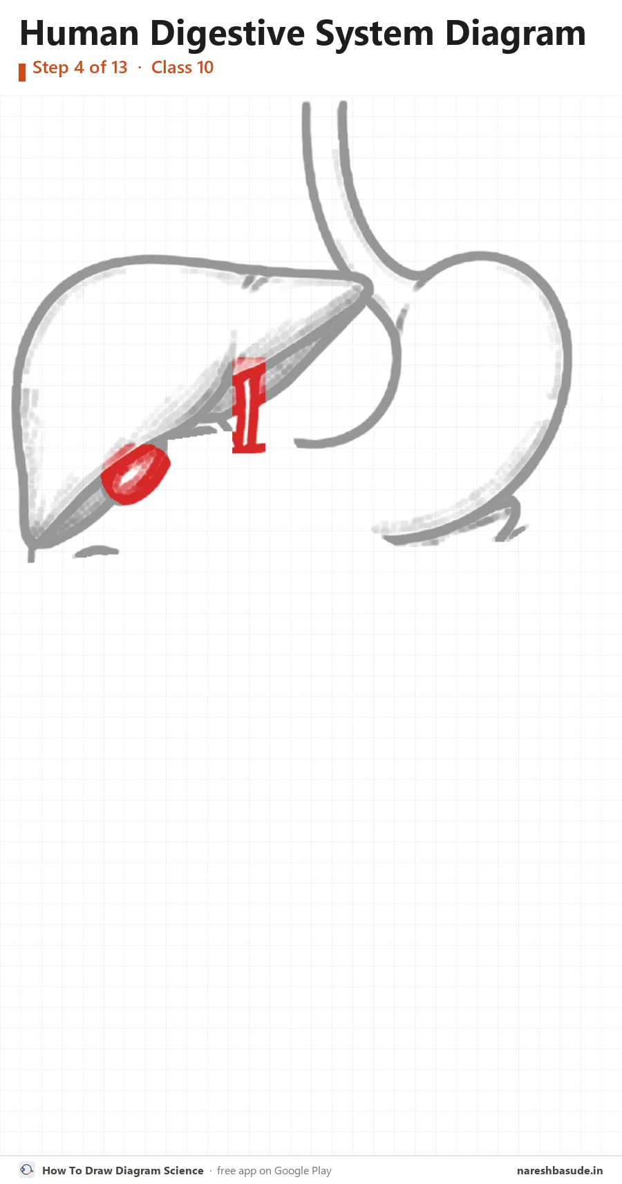
Gall bladder
Under the liver draw the small gall bladder, with a thin bile duct running down towards the intestine.
Parts drawn: Gall bladder
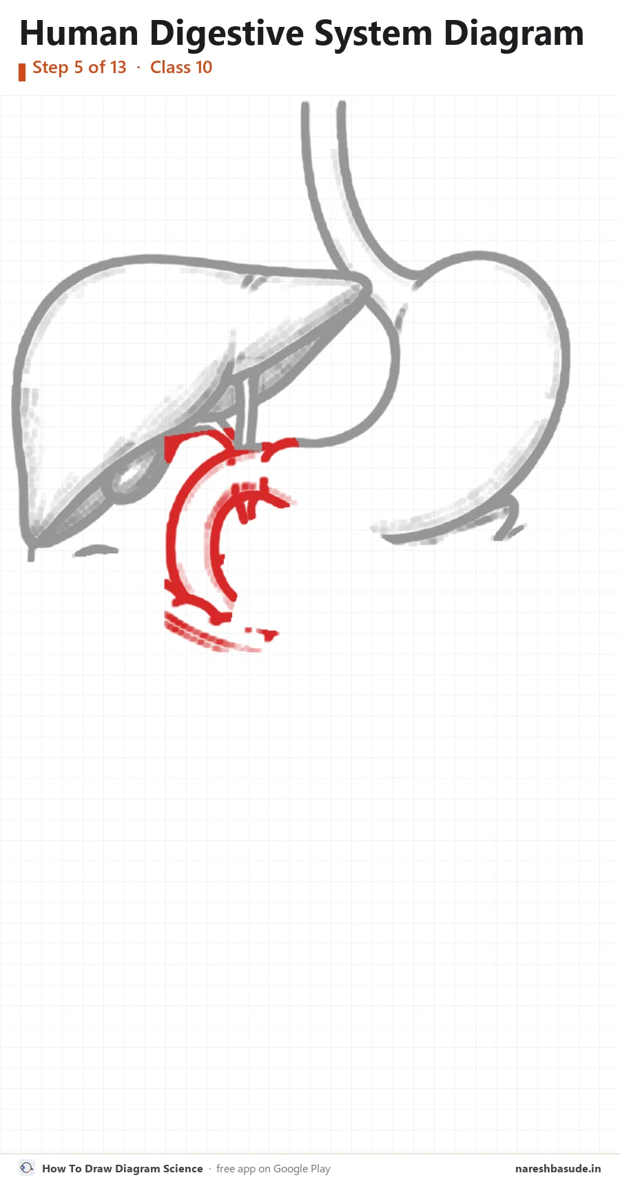
Duodenum
From the lower end of the stomach, draw a C-shaped tube curving down: the first part of the small intestine.
Parts drawn: Duodenum
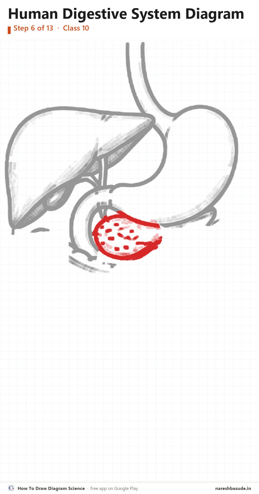
Pancreas
Inside the curve of the C, draw the pancreas: a soft leaf-shaped gland under the stomach.
Parts drawn: Pancreas
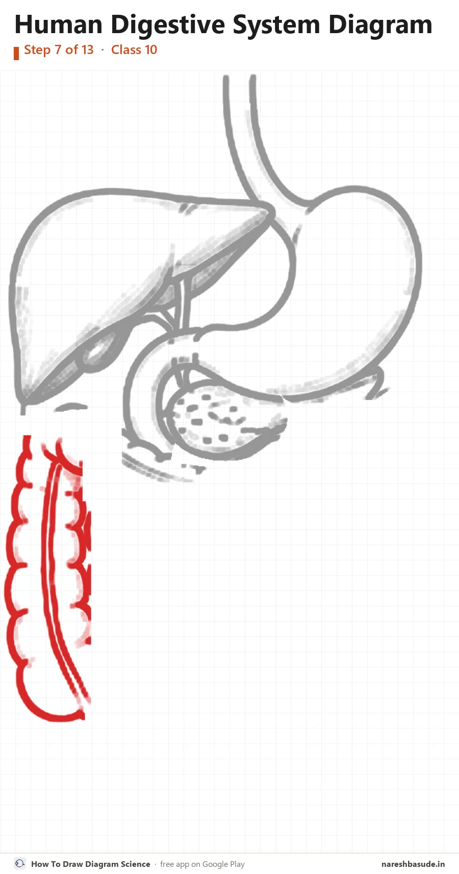
Large intestine: rising on the left
Draw the large intestine as a wide bumpy tube. Start low on the left and go straight up.
Parts drawn: Large intestine
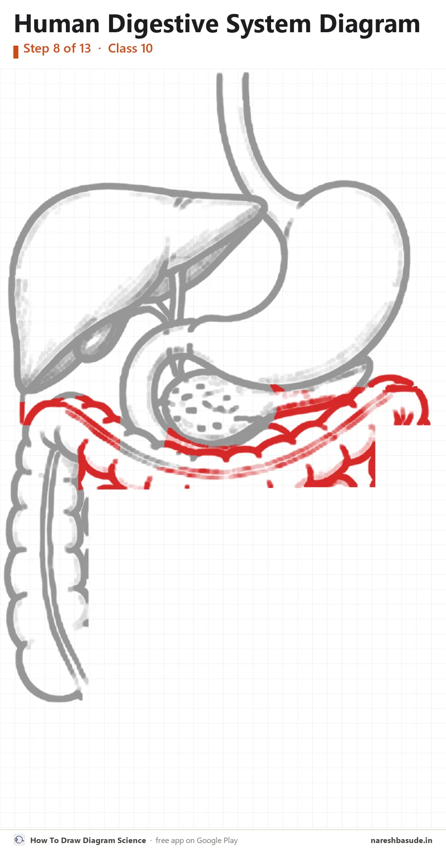
Large intestine: across the top
Carry the bumpy tube across the top, just under the stomach and pancreas.
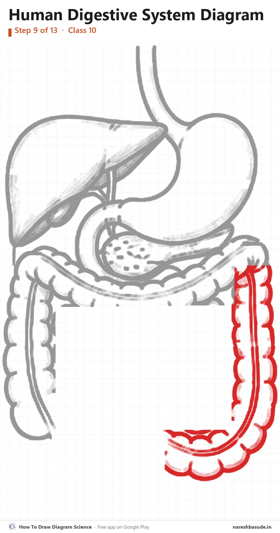
Large intestine: down the right
Bring it down the right side and curve it in towards the middle at the bottom.
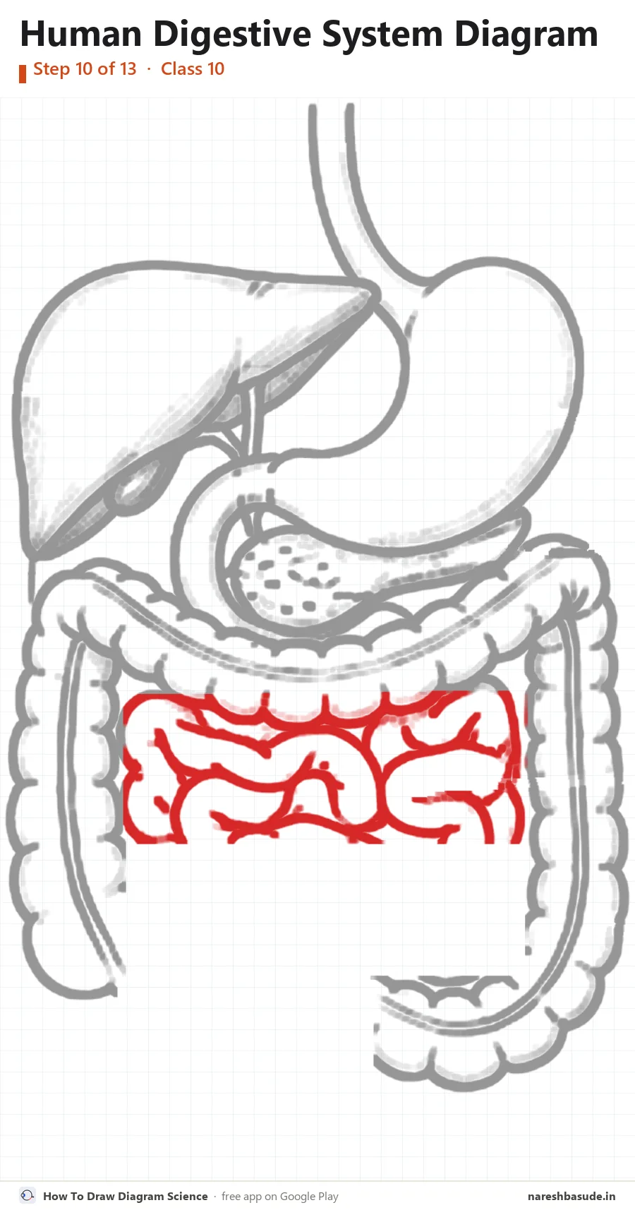
Small intestine: upper coils
Fill the middle with the small intestine: a long, narrow tube folded into tight coils.
Parts drawn: Small intestine
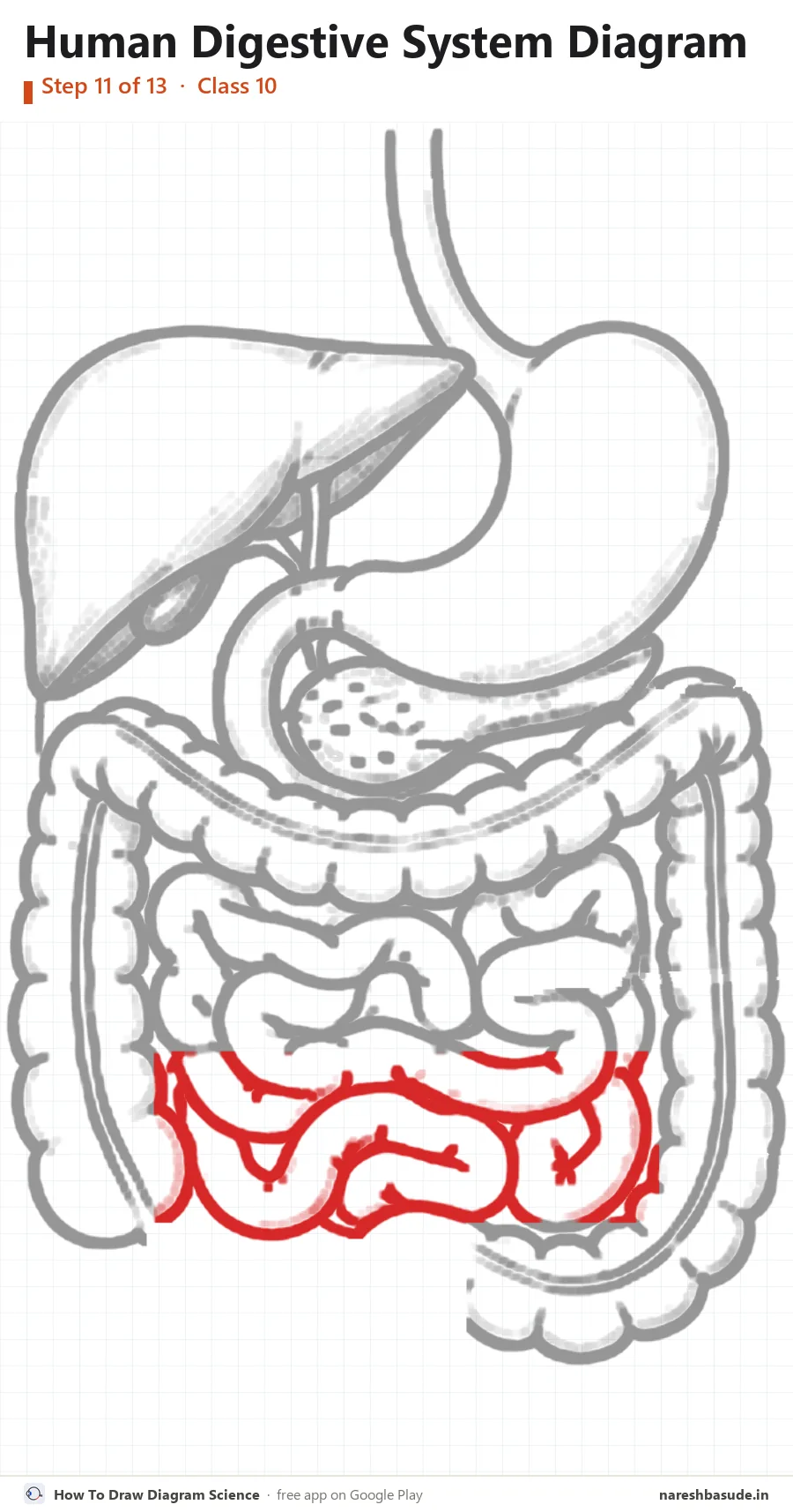
Small intestine: lower coils
Keep coiling the tube down to the bottom of the space.
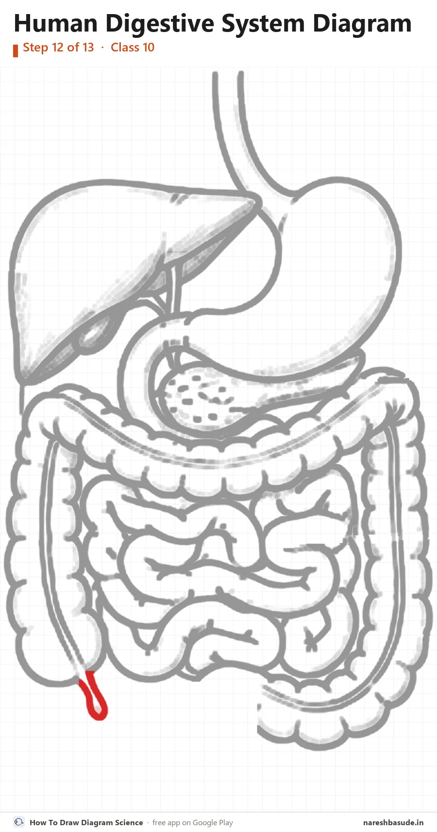
Appendix
At the bottom of the left side of the large intestine, add the small finger-like appendix.
Parts drawn: Appendix
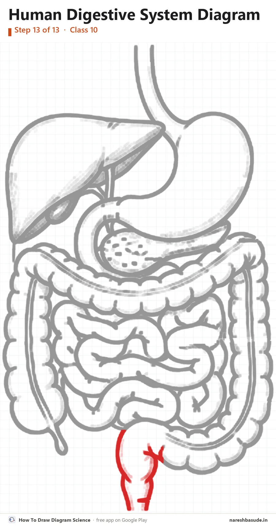
Rectum and anus
In the middle at the bottom, draw the short straight tube going down: the rectum, ending at the anus.
Parts drawn: Rectum, Anus

Parts to label (11) and what each one does
- Liver The largest gland in the body. It makes bile, which breaks fats into small droplets.
- Gall bladder A small sac under the liver that stores bile and releases it into the duodenum.
- Duodenum The first part of the small intestine. Bile and pancreatic juice enter here.
- Large intestine Takes back extra water from the undigested food.
- Appendix A small, finger-shaped pouch where the small and large intestines meet. It has no known role in human digestion.
- Rectum Stores the waste (faeces) until it is passed out.
- Oesophagus The food pipe. Its muscles push food down from the mouth to the stomach (peristalsis).
- Stomach A muscular bag that churns food with gastric juice (hydrochloric acid, pepsin and mucus) and starts protein digestion.
- Pancreas A gland below the stomach. Its pancreatic juice has enzymes, such as trypsin and lipase, that digest proteins and fats.
- Small intestine The longest part of the canal. Digestion is completed here, and digested food is absorbed through its villi into the blood.
- Anus The opening through which waste leaves the body. A ring of muscle (the anal sphincter) controls it.
Write each name on the side of the diagram, joined to its part with a straight line. Keep the lines from crossing.
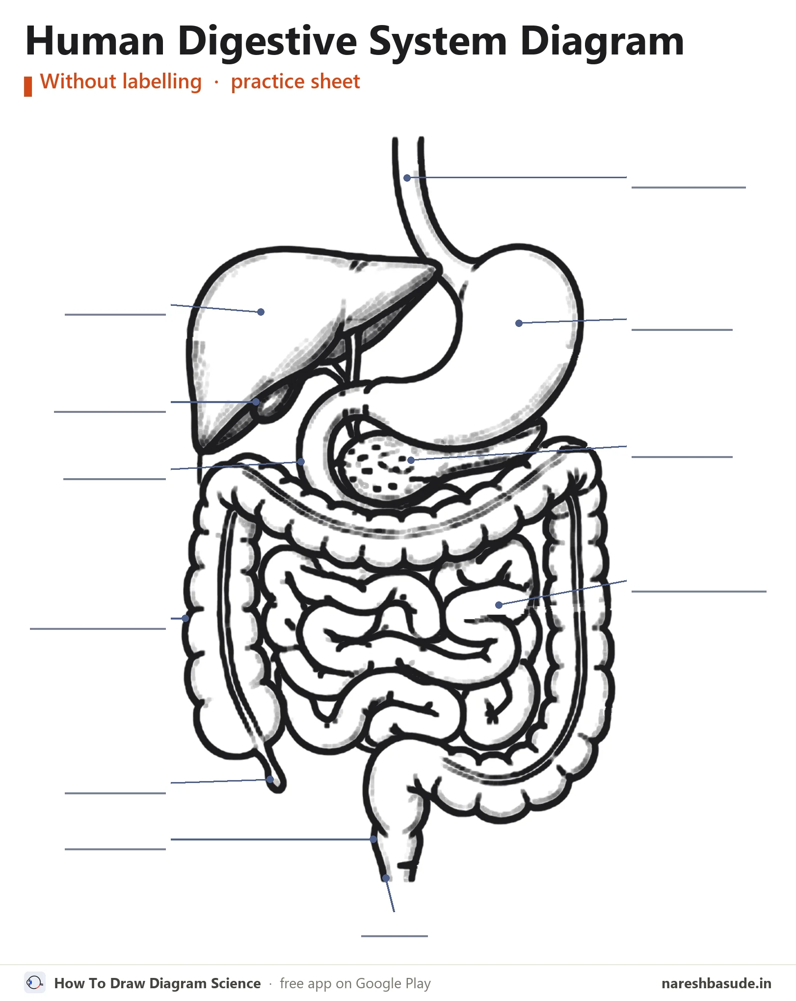
Human Digestive System diagram without labelling (practice sheet)
The same diagram with the label lines but no names (an unlabelled diagram). Write each part's name on its line, then check your answers against the labelled diagram at the top.
Download free PDF (A4, 3 pages)
Page 1: the labelled diagram. Page 2: this practice sheet with a word bank of all 11 parts. Page 3: all 13 drawing steps on one page.
Exam tips
- Begin with the oesophagus and the J-shaped stomach, then add the liver on the other side.
- Draw the large intestine as a frame first, then fill the middle with the coils of the small intestine.
- Make the small intestine narrow and tightly coiled, and the large intestine wide and bumpy.
Common mistakes
- Putting the liver on the same side as the stomach.
- Swapping the labels for the small and large intestine.
- Leaving out the gall bladder, the pancreas or the appendix.
Exam questions on the human digestive system diagram
QuestionDraw a diagram of the human alimentary canal and label: gall bladder, liver and pancreas.
The liver is the large organ on your left, just under the diaphragm, with the gall bladder tucked under it. The pancreas lies below the stomach, in the loop of the duodenum. Draw the rest of the canal, from the oesophagus to the anus, around them.
QuestionWhat is the role of bile in digestion?
Bile is made by the liver and stored in the gall bladder. In the small intestine it makes the food alkaline so the pancreatic enzymes can work, and it breaks fats into small droplets (emulsification).
Questions students ask
Which is the largest gland in the human body?
The liver.
Where is bile stored?
In the gall bladder, which sits under the liver.
Where is digestion completed?
In the small intestine, which is also where the digested food is absorbed.
Practise it on your phone
These steps come straight from the free Android app How To Draw Diagram Science. In the app you can view the human digestive system with every part named, learn to draw it step by step on a numbered grid, take a quiz on the parts and label it yourself.


