How to Draw and Label a Human Eye Diagram, Step by Step
The human eye diagram is part of Class 10 Physics (The Human Eye and the Colourful World). It shows how light gets in through the cornea, passes through the pupil and the lens, and forms an image on the retina. The steps below build the eye from the outside in.
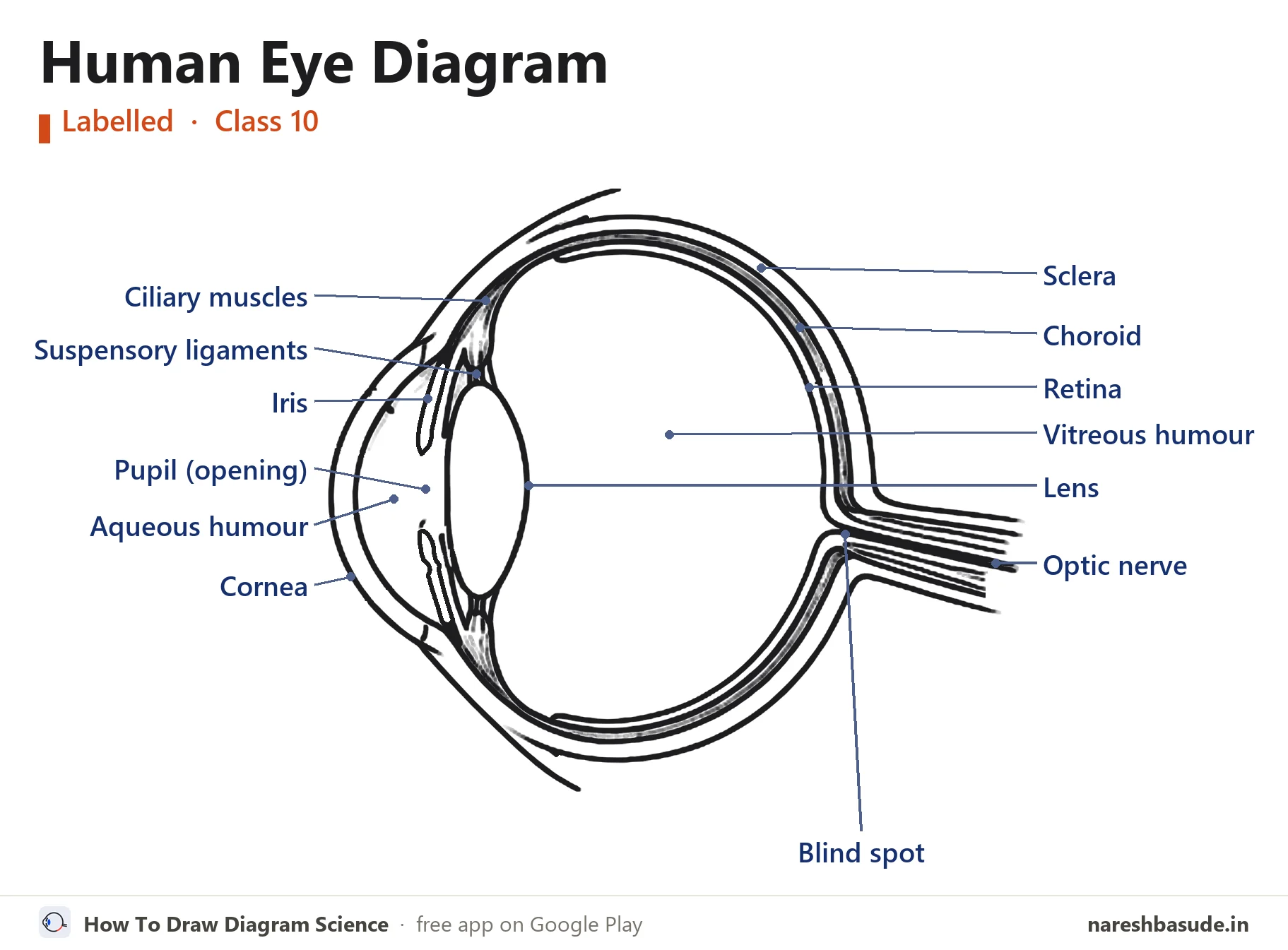
The finished human eye diagram with all 13 parts labelled. The steps below build it up one part at a time.
What the human eye does
Light enters through the cornea, passes the pupil (an opening in the iris) and is focused by the lens onto the retina. The optic nerve carries the signal to the brain. The ciliary muscles change the thickness of the lens so the eye can focus on near and far objects.
Draw it in 12 steps
Use a sharp pencil and draw lightly at first. In every picture, red is what to draw in that step and grey is what you have already drawn.
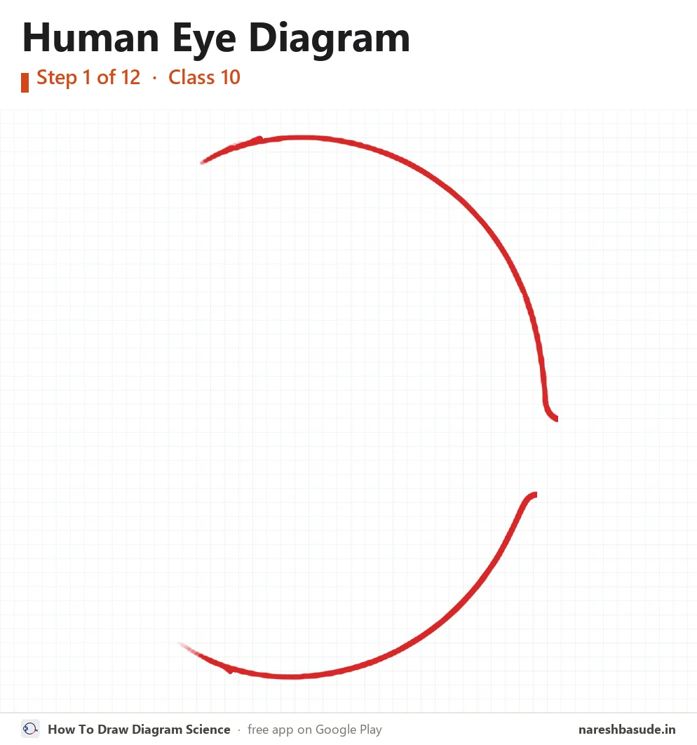
Outer circle
Draw a big round circle for the eyeball, but leave the front (left side) open: the cornea goes there.
Parts drawn: Sclera
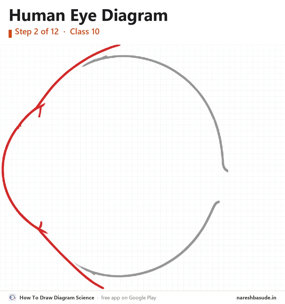
Cornea, outer line
Close the front with a curve that bulges out to the left. This clear window is the cornea.
Parts drawn: Cornea
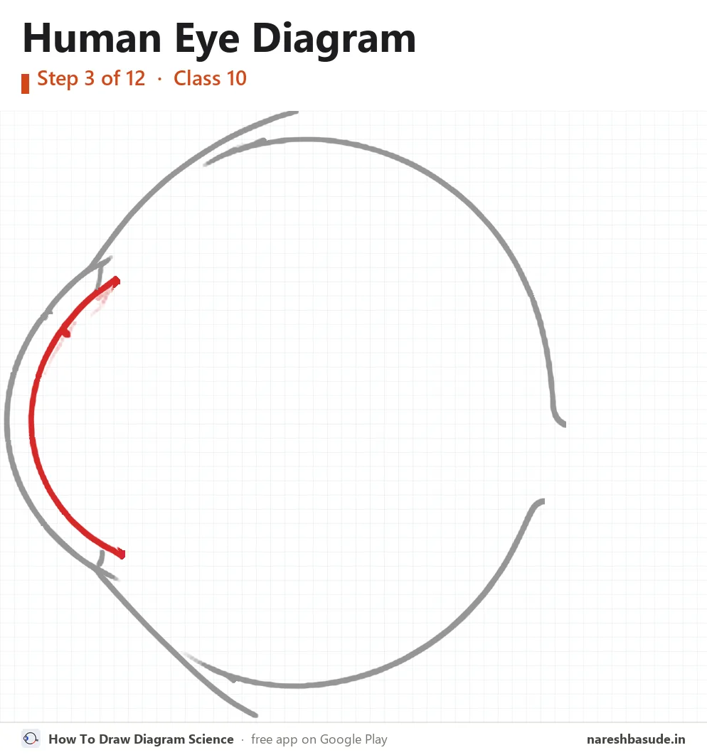
Cornea, inner line
Draw a second curve just inside the first one, the same shape. The cornea is the thick layer between the two lines.
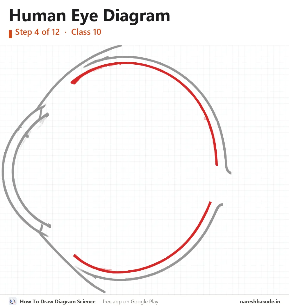
Sclera
Just inside the big circle, draw a second circle. The white band between the two lines is the sclera, the tough outer coat.
Parts drawn: Sclera
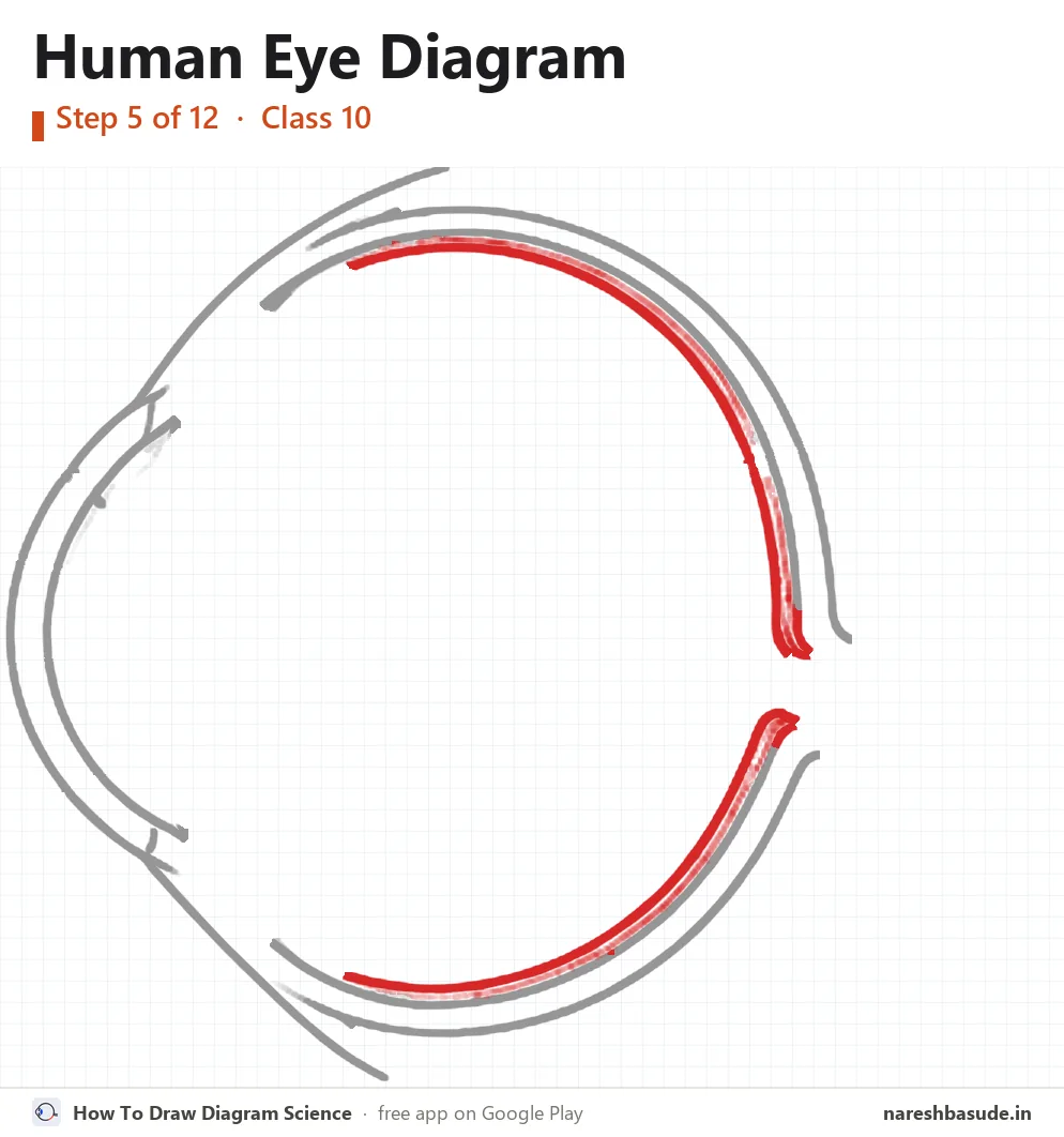
Choroid
Inside the sclera draw the next thin layer, the choroid, following the same curve.
Parts drawn: Choroid
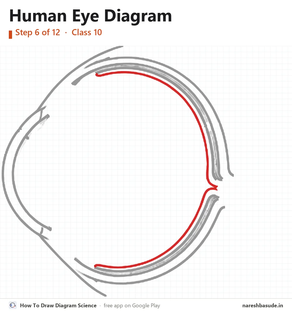
Retina
Draw the innermost line all the way round: this is the retina, where the picture forms.
Parts drawn: Retina
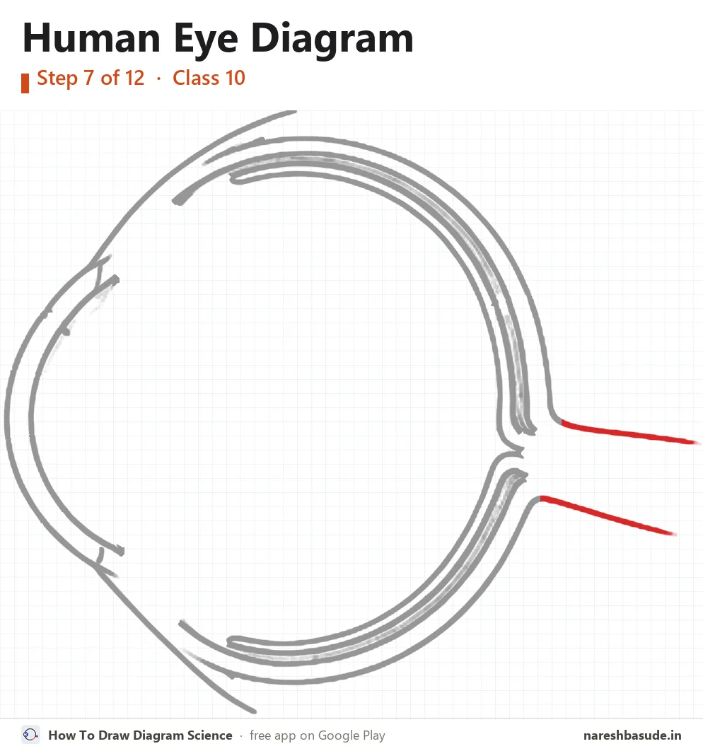
Optic nerve tube
At the back (right side) let the outer lines run out into a short tube.
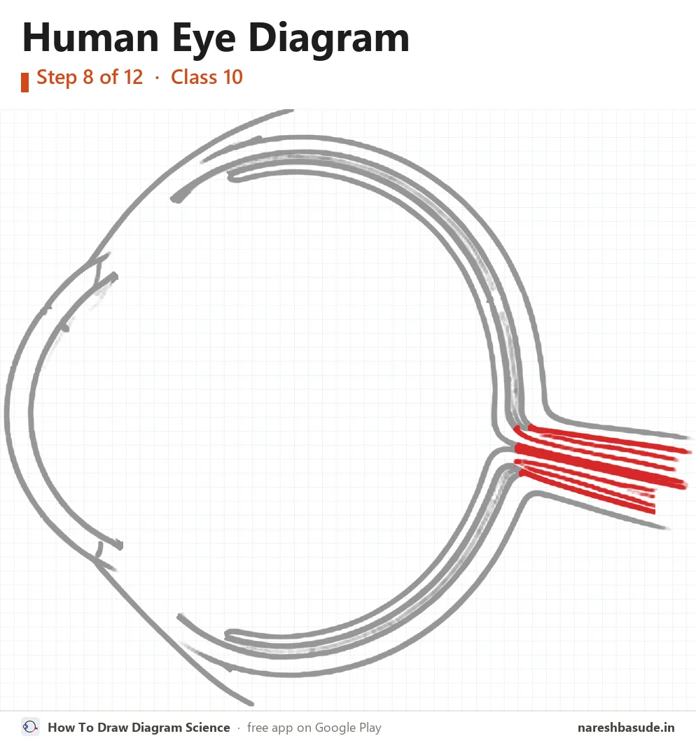
Optic nerve
Fill the tube with long lines: this is the optic nerve. Where it leaves the retina there is no sight: the blind spot.
Parts drawn: Optic nerve, Blind spot
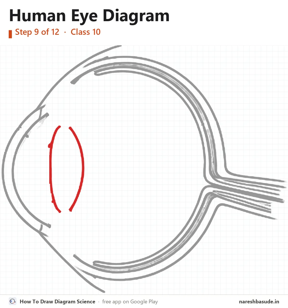
Lens
Behind the cornea draw the lens: a tall oval that bulges on both sides.
Parts drawn: Lens, Vitreous humour
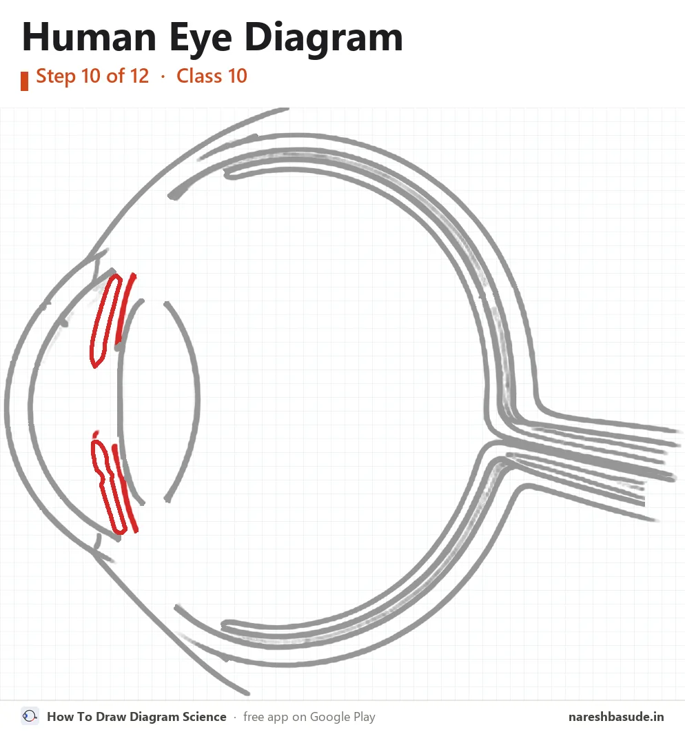
Iris and pupil
In front of the lens draw the iris as two flaps, one above and one below. Leave a gap between their tips: that gap is the pupil, a hole that lets light in.
Parts drawn: Iris, Pupil, Aqueous humour
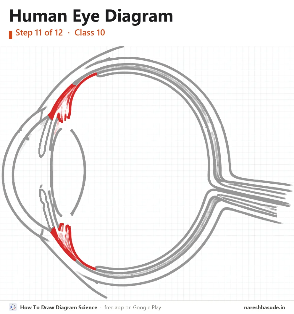
Ciliary muscles
Above and below the lens draw the ciliary muscles: the shaded fan shapes joined to the wall.
Parts drawn: Ciliary muscles
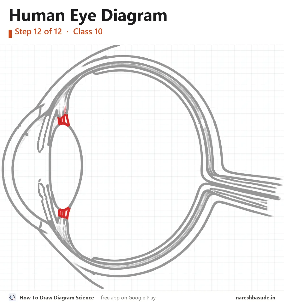
Suspensory ligaments
Join the ciliary muscles to the top and bottom of the lens with a few short threads: the suspensory ligaments.
Parts drawn: Suspensory ligaments

Parts to label (13) and what each one does
- Ciliary muscles Muscles around the lens that make it thicker or thinner so the eye can focus (accommodation).
- Suspensory ligaments Fibres that hold the lens in place and join it to the ciliary muscles.
- Iris The coloured, muscular ring behind the cornea. It controls the size of the pupil.
- Pupil (opening) The hole in the middle of the iris that lets light in. It gets smaller in bright light and wider in dim light.
- Aqueous humour Watery fluid between the cornea and the lens that keeps the front of the eye in shape.
- Cornea The clear front window of the eye. Most of the bending (refraction) of light happens here.
- Sclera The tough white outer coat that protects the eyeball.
- Choroid The dark layer under the sclera. Its blood vessels feed the eye, and its colour stops light from scattering inside.
- Retina The light-sensitive screen at the back of the eye. Its rods and cones turn light into nerve signals.
- Vitreous humour Clear jelly that fills the eyeball between the lens and the retina and keeps it round.
- Lens A clear, flexible convex lens that focuses light on the retina. It changes shape to see near and far objects.
- Optic nerve Carries the signals from the retina to the brain.
- Blind spot The point where the optic nerve leaves the eye. It has no rods or cones, so no image forms there.
Write each name on the side of the diagram, joined to its part with a straight line. Keep the lines from crossing.
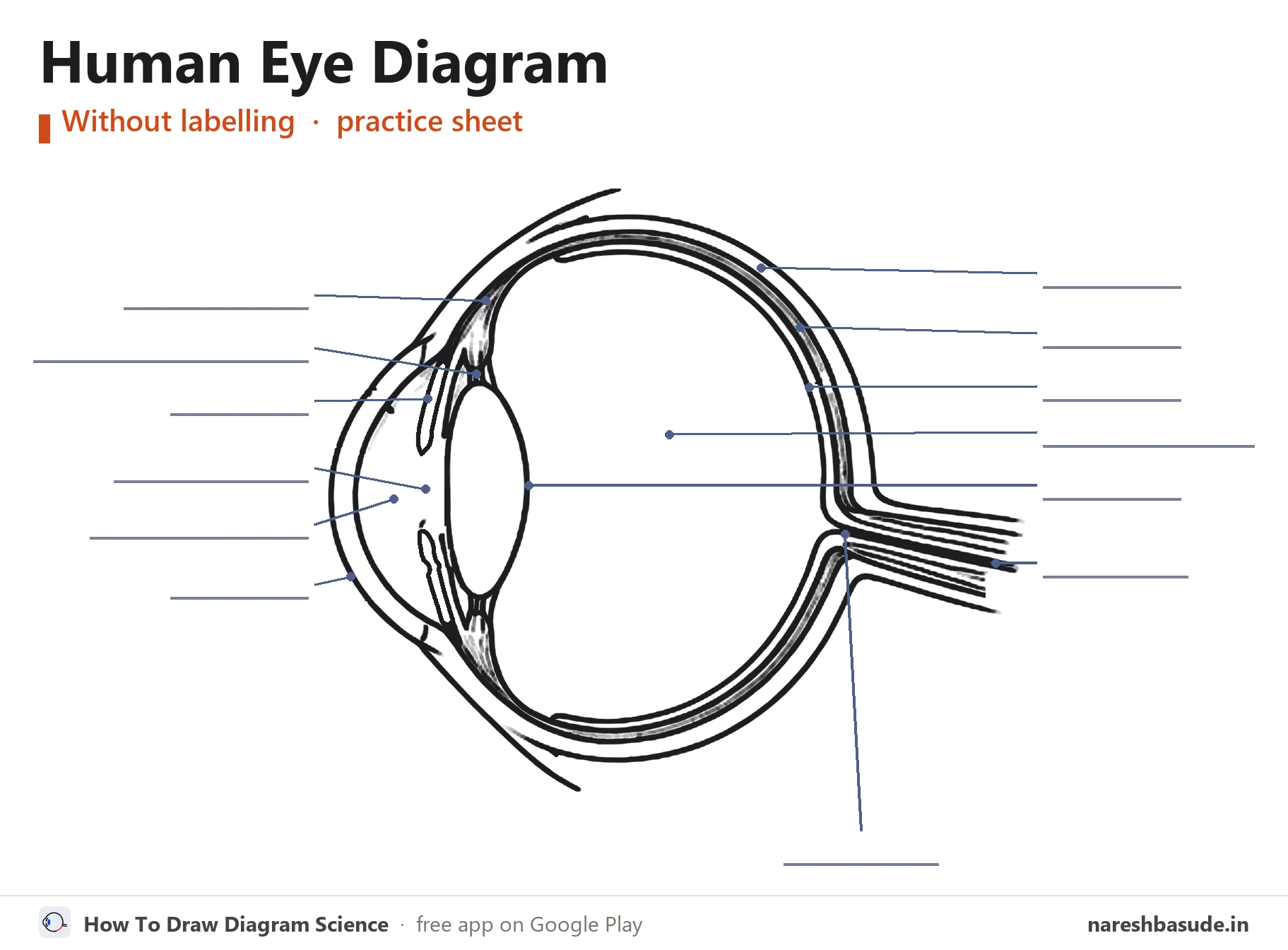
Human Eye diagram without labelling (practice sheet)
The same diagram with the label lines but no names (an unlabelled diagram). Write each part's name on its line, then check your answers against the labelled diagram at the top.
Download free PDF (A4, 3 pages)
Page 1: the labelled diagram. Page 2: this practice sheet with a word bank of all 13 parts. Page 3: all 12 drawing steps on one page.
Exam tips
- Make the cornea bulge out a little more than the rest of the eyeball.
- Draw the lens as a tall oval that bulges on both sides, just behind the iris.
- Join the ciliary muscles to the top and bottom of the lens with the suspensory ligaments.
- Show the optic nerve leaving from the back, and mark the blind spot where it leaves.
Common mistakes
- Drawing the lens in front of the iris instead of behind it.
- Mixing up the aqueous humour (in front of the lens) and the vitreous humour (behind it).
- Treating the pupil as a solid part. It is an opening in the iris that lets light in.
Exam questions on the human eye diagram
QuestionDraw a labelled diagram of the human eye and label: cornea, iris, pupil, ciliary muscles and retina.
The cornea is the bulging clear layer at the front. The iris is the ring just behind it, and the pupil is the gap in the middle of the iris. The ciliary muscles sit around the lens and hold it through the suspensory ligaments. The retina lines the back of the eye.
QuestionWhat is the power of accommodation of the eye?
It is the eye lens's ability to change its focal length so that both near and far objects are seen clearly. The ciliary muscles do this by making the lens thicker for near objects and thinner for far ones.
Questions students ask
Where is the image formed in the human eye?
On the retina, the light-sensitive layer at the back of the eye.
What is the blind spot?
The point where the optic nerve leaves the eye. It has no light-sensitive cells, so no image forms there.
What controls the size of the pupil?
The iris. It makes the pupil smaller in bright light and larger in dim light.
Practise it on your phone
These steps come straight from the free Android app How To Draw Diagram Science. In the app you can view the human eye with every part named, learn to draw it step by step on a numbered grid, take a quiz on the parts and label it yourself.


