Human Reproduction
Class 12 Biology notes, explained simply
How does a new human life begin? This chapter follows the whole story in order: where sperm and eggs are made, how they meet, how the baby grows for nine months, and how it is born. Plain words, diagrams, memory tricks, exam tips and a 10-question self-check.
In short: The testes make sperm and the ovaries make eggs. When a sperm and an egg meet, a new life starts. It grows inside the mother for 9 months and becomes a baby.
Prefer Hindi? Read this chapter in Hindi notes (Hinglish).
The whole chapter in 30 seconds
- Gametes are madeThe testes make sperm (spermatogenesis); the ovaries make eggs (oogenesis). Both have 23 chromosomes.
- A monthly clockIn the menstrual cycle, an LH surge releases one egg around day 14, and the uterus lining gets ready.
- FertilisationSperm meets the egg in the ampulla of the fallopian tube. The zygote has 46 chromosomes, and the sperm decides boy or girl.
- Implantation and pregnancyThe zygote becomes a blastocyst, sinks into the endometrium, and the placenta feeds the baby for 9 months.
- Birth and milkOxytocin drives the uterus to push the baby out (parturition). The first milk, colostrum, is full of antibodies.
Remember: sperm formation goes on into old age, but egg release stops around 50 (menopause).
Contents14 parts · quiz · FAQ
- The whole story of new life
- Testis: the sperm factory
- The path of the sperm
- The ovary and the fallopian tube
- The uterus and the other parts
- How sperm are made, and the hormones
- The structure of a sperm, and semen
- How an egg is made
- Menstrual cycle: the 28-day clock
- Fertilisation and boy or girl
- Zygote to blastocyst, and implantation
- Pregnancy: placenta and the baby's growth
- Birth and the mother's milk
- Quick revision
- Self-check quiz
- Words to know
- Questions students ask
Dotted words are explained: tap one to see what it means.
1How does new life begin in a human?
In short: the father's body makes sperm, and the mother's body makes an ovum. When the two meet, a new life starts. It grows inside the mother for 9 months and becomes a baby.
Guess firstA hen lays an egg, and the chick grows inside the egg. Where does a human baby grow?
Inside the mother's body. The baby grows there and is born alive. The exam name for this is below.
What is similar to Chapter 1?
In Chapter 1 (Sexual Reproduction in Flowering Plants) you read the flower's story. The male cell and the female cell met, and a seed was made. In humans the idea is the same.
The father's male gamete is called a sperm. The mother's female gamete is called an ovum (egg). When they meet, a zygote is formed, and the zygote becomes a baby.
A baby needs both a male and a female cell. So humans do sexual reproduction.
What does viviparous mean?
Hens, sparrows and snakes lay eggs. The young one grows outside the mother, inside the egg. Such animals are called oviparous.
Humans, cows, dogs and cats give birth to live young ones. The young one grows inside the mother. Such animals are called viviparous.
Trick: Oviparous = ovum (egg), so they lay eggs. Viviparous: "vivi" means alive, so they give birth to live young ones.
The game of 23 and 46
In Chapter 1 you read that the nucleus of a cell holds information. This information is written on tiny threads called chromosomes.
Every normal human cell has 46 chromosomes: 23 from the mother and 23 from the father. We call this 2n.
A sperm and an ovum have only half, which is 23 chromosomes (n). When they meet, 23 + 23 = 46. The zygote again has the full 46.
Remember: sperm = 23, ovum = 23, zygote = 46. Gametes are n, the zygote is 2n.
What are the 6 steps of the story?
The whole chapter is a story of 6 steps. Here is each name in one line. Later, each step gets its own part.
- Gametogenesis: making the sperm and the ovum
- Insemination: the sperm reaching inside the female's body
- Fertilisation: the sperm and ovum meeting, and the zygote forming
- Implantation: the growing baby attaching to the wall of the uterus (garbhashaya)
- Gestation: the baby growing inside the mother for 9 months
- Parturition: the birth of the baby
Trick: remember the 6 jobs, and the order comes by itself: made (gametogenesis), reached (insemination), met (fertilisation), attached (implantation), grew (gestation), born (parturition).
When does all this begin?
This does not happen in childhood. It begins when the body starts to grow up and change. This age is called puberty.
RiyaSir, so do men and women both make gametes all their life?
SirNo, there is a big difference here! In men, sperm keep being made until old age.
SirIn women, making ova stops at around 50 years of age. You will read about this in detail in Part 9.
This chapter uses the names of some body parts. This is normal biology, like the names of parts of the heart or the stomach. Doctors and NCERT use these plain names too.
Don't mix them up: fertilisation (the meeting) and implantation (attaching to the uterus) are different steps. Fertilisation comes first, and implantation comes later.
Hindi medium? See the Hindi names (tap to open)
| English | Hindi name | Meaning of the pieces |
|---|---|---|
| Sperm | शुक्राणु | शुक्र (male fluid) + अणु (tiny particle) |
| Ovum | अंडाणु | अंड (egg) + अणु (tiny particle) |
| Zygote | युग्मनज | युग्म (pair) + ज (born from) |
| Viviparous | जरायुज | Gives birth to live young ones |
| Gametogenesis | युग्मकजनन | युग्मक (gamete) + जनन (making) |
| Fertilisation | निषेचन | The meeting of a male and a female gamete |
| Parturition | प्रसव | The birth of the baby |
| Puberty | यौवनारंभ | यौवन (youth) + आरंभ (start) |
Revise: 6 cards (tap one to flip it)
Sperm / Ovum
The male gamete / the female gamete (egg). Each has 23 chromosomes.
Zygote
The cell formed when a sperm and an ovum meet. 46 chromosomes (2n).
Viviparous
Gives birth to live young ones, like humans and cows.
Oviparous
Lays eggs, like hens and snakes.
6 steps
Gametogenesis, insemination, fertilisation, implantation, gestation, parturition.
Puberty
The age when the body grows up and gametes start to be made.
Check yourself
How many chromosomes does a human zygote have?
46. That is 23 from the sperm plus 23 from the ovum.
What is it called when the baby attaches to the wall of the uterus?
Implantation. Fertilisation comes first, and parturition (birth) comes last.
What kind of animal is a human?
Viviparous: the baby grows inside the mother and is born alive.
Now let us start the story from the father's side: where is sperm made?
A small "factory" hangs outside the body, because it needs to stay a little cool. Why? In the next part.
2What is inside the testis?
In short: sperm are made in two testes. The testes hang outside the belly in a bag, because making sperm needs a slightly lower temperature. Inside each testis there are very long, coiled tubes, and sperm are made inside them.
Guess firstWhy are the testes not inside the belly? Why do they hang outside in a bag?
It needs to be cooler! The heat inside the belly is too much for making sperm. See below how much cooler.
What is in the male reproductive system?
This system sits in the lowest part of the belly, called the pelvis. It has 4 things: 2 testes, the tubes (ducts) that carry sperm, some helper glands, and the outer part, the penis.
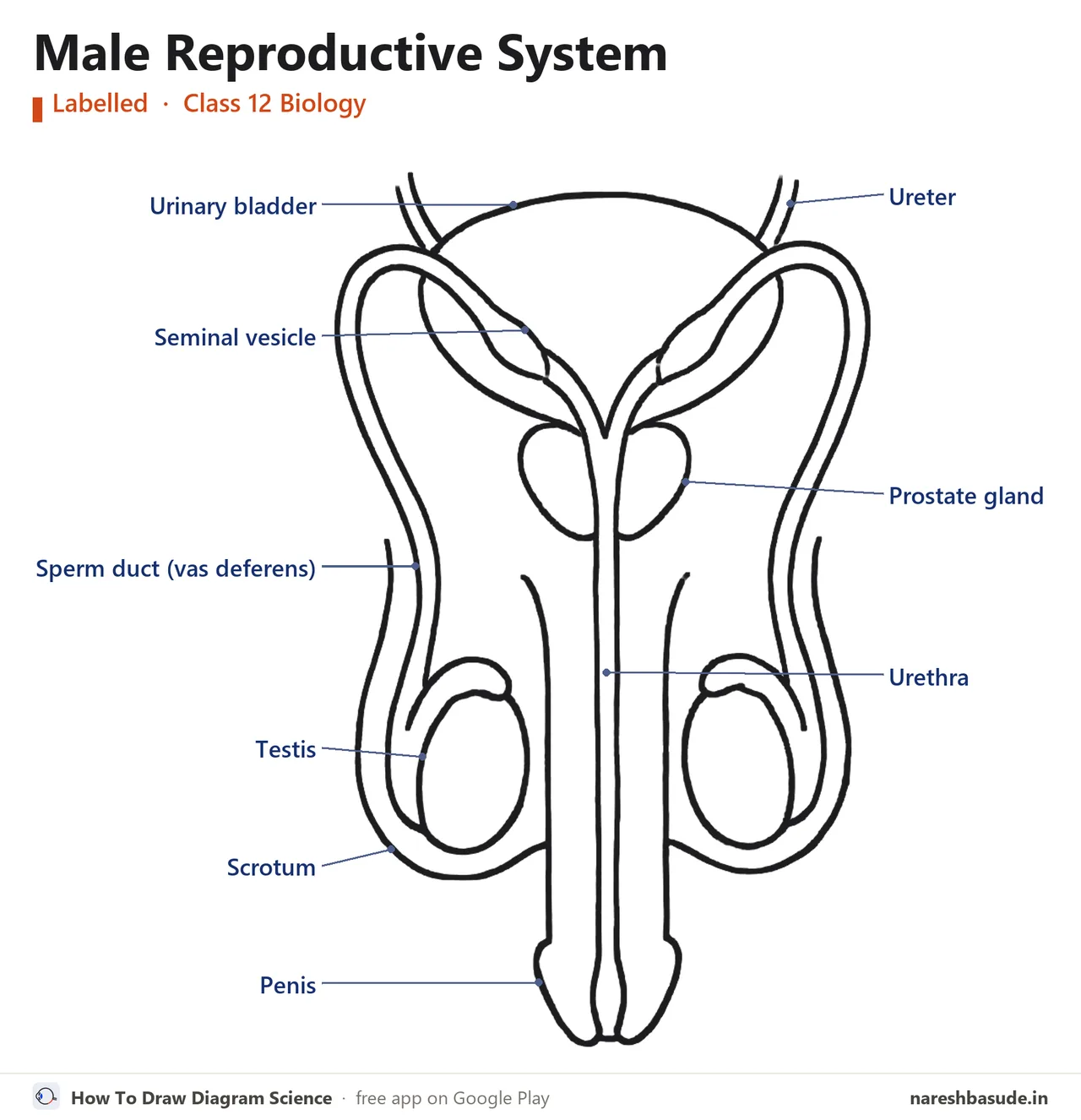
Why do the testes hang outside?
The testes are not inside the belly. They hang in a bag of skin called the scrotum.
The scrotum keeps the testes 2 to 2.5 °C cooler than the temperature inside the body. Sperm can only be made at this cooler temperature.
In a grown man, each testis is oval. It is about 4 to 5 cm long and 2 to 3 cm wide, with a tough cover on top.
What is inside the testis?
Inside the testis there are about 250 small rooms. These rooms are called testicular lobules.
Each room has 1 to 3 very long tubes, coiled round and round like noodles on a plate. They are called seminiferous tubules. Sperm are made inside these tubes.
- Testis
- About 250 rooms (testicular lobules)
- 1-3 coiled tubes in each room (seminiferous tubules)
- Sperm are made inside the tubes
Who is inside the tube?
The inner wall of each seminiferous tubule has two kinds of cells.
- Male germ cells, called spermatogonia. They divide (by meiosis) and finally become sperm.
- Sertoli cells: they feed the growing germ cells, like a nanny feeding children.
What is outside the tube?
The empty space between the tubes is called the interstitial space. It has small blood vessels and some special cells.
These are the Leydig cells (or interstitial cells). They make male hormones.
These hormones are called androgens. There are also some immune cells here. They protect the body from disease.
RiyaSir, so do the Leydig cells also make sperm?
SirNo! This is the most common mistake. Sperm are made only inside the tubes, from the germ cells.
SirLeydig cells sit outside the tubes, and they only make hormones (androgens).
Don't mix them up: Sertoli cells are inside the tube and give food. Leydig cells are outside the tube and make androgens.
Trick: Sertoli = Serve (they serve food). Leydig = Lie outside (they stay outside) and make hormones.
Hindi medium? See the Hindi names (tap to open)
| English | Hindi name | Meaning of the pieces |
|---|---|---|
| Testis | वृषण | The gland that makes sperm |
| Scrotum | वृषणकोश | वृषण + कोश (bag) |
| Testicular lobules | वृषण पालिकाएँ | वृषण + पालिका (small part) |
| Seminiferous tubules | शुक्रजनक नलिकाएँ | शुक्र + जनक (maker) + नलिका (tube) |
| Spermatogonia | शुक्राणुजन | The first cells that make sperm |
| Sertoli cells | सर्टोली कोशिकाएँ | Just the English name |
| Leydig cells | लीडिग कोशिकाएँ | Just the English name |
| Androgens | एंड्रोजन | Male hormones |
Revise: 7 cards (tap one to flip it)
Scrotum
The bag of the testes. It keeps them 2-2.5 °C cooler than the body.
Testicular lobules
About 250 rooms in the testis.
Seminiferous tubules
1-3 coiled tubes in each lobule. Sperm are made here.
Spermatogonia
Male germ cells in the tube wall. Sperm are made from them.
Sertoli cells
Inside the tube. They feed the germ cells.
Leydig cells
Outside the tube (interstitial space). They make androgens.
Size of the testis
Oval, 4-5 cm long, 2-3 cm wide.
Check yourself
Who makes androgens?
Leydig cells, which sit outside the tubes in the interstitial space.
How many seminiferous tubules does one lobule of the testis have?
1 to 3. The lobules (rooms) number about 250.
By how much does the scrotum keep the testes cooler than the body?
2 to 2.5 °C cooler. This is needed to make sperm.
The sperm are made, but they are still inside the tubes. How will they get outside?
Which tubes are on the way, and which glands add "food and water" to them? In the next part.
3By which path do sperm leave the testis?
In short: sperm are made in the seminiferous tubules and travel through several tubes to the urethra. The urethra opens outside through the penis. These tubes also store the sperm and carry them forward.
Guess firstDo urine (peshaab) and sperm come out through the same tube?
Yes! The urethra starts at the bladder, and the sperm tube also joins it at the end.
The journey of the sperm: which tubes?
Think of a train route. The sperm pass through many "stations" before they come out.
Read this together with the diagram in Part 2, and find the vas deferens and the urethra.
- Seminiferous tubules: sperm are made here
- Rete testis: all the tubes join in a network here
- Vasa efferentia: small tubes that leave the testis
- Epididymis: a long coiled tube stuck to the back of the testis
- Vas deferens: a long tube that goes up into the belly, passes over the urinary bladder, and comes down
- Ejaculatory duct: here the duct of a gland (the seminal vesicle) joins the vas deferens
- Urethra: the last tube, which goes out through the penis
NCERT calls 4 of these tubes the male accessory ducts: the rete testis, vasa efferentia, epididymis and vas deferens.
Trick, 2 + 3 + 2: 2 inside the testis (seminiferous tubules, rete testis). 3 after leaving the testis (vasa efferentia, epididymis, vas deferens). The last 2 (ejaculatory duct, urethra).
The urethra and the penis
The urethra starts at the urinary bladder and opens outside by passing through the penis. Its outer opening is called the urethral meatus.
The penis is the male's outer reproductive part (external genitalia). It has a special tissue that helps it become firm (erection). This helps the sperm reach inside the female, which is called insemination.
The swollen front part of the penis is called the glans penis. It is covered by a loose fold of skin called the foreskin.
RiyaSir, what do the sperm do when they stop in the epididymis?
SirThese tubes store the sperm for some time. Their fluid helps the sperm become mature and able to swim. The details are in Part 7.
Don't mix them up: the vas deferens does not open straight into the urethra. First it joins the duct of the seminal vesicle to make the ejaculatory duct, and that opens into the urethra.
Hindi medium? See the Hindi names (tap to open)
| English | Hindi name | Meaning of the pieces |
|---|---|---|
| Rete testis | वृषण जालिकाएँ | वृषण + जालिका (network) |
| Vasa efferentia | शुक्रवाहिकाएँ | शुक्र + वाहिका (carrying tube) |
| Epididymis | अधिवृषण | अधि (on, beside) + वृषण |
| Vas deferens | शुक्रवाहक | शुक्र + वाहक (carrier) |
| Ejaculatory duct | स्खलनीय वाहिनी | स्खलन (coming out) + वाहिनी |
| Urethra | मूत्रमार्ग | मूत्र (urine) + मार्ग (path) |
| Penis | शिश्न | The male's outer reproductive part |
Revise: 6 cards (tap one to flip it)
The path
Seminiferous tubules, rete testis, vasa efferentia, epididymis, vas deferens, ejaculatory duct, urethra.
Epididymis
A coiled tube on the back of the testis. The vas deferens starts from it.
Vas deferens
Goes up into the belly and passes over the bladder.
Ejaculatory duct
Vas deferens + duct of the seminal vesicle. Opens into the urethra.
Urethra
From the bladder, out through the penis. Outer opening: urethral meatus.
Glans penis
The front part of the penis, covered by the foreskin.
Check yourself
What do the vas deferens and the duct of the seminal vesicle make when they join?
The ejaculatory duct, which opens into the urethra.
Which tube is not inside the testis?
The vas deferens is outside the testis and goes up into the belly. Trick: 2 inside, 3 after leaving, last 2.
Where does the urethra start?
At the urinary bladder. It passes through the penis and opens outside at the urethral meatus.
Now let us go to the mother's side. Where is the egg made, and by which path does it go forward?
There, a part like the "fingers of a hand" catches the egg. In the next part.
4Where is the egg made, and by which path does it go forward?
In short: the egg (ovum) is made in two ovaries. The egg that leaves the ovary goes into a thin tube called the fallopian tube. Finger-like parts at the end of the tube catch the egg and take it inside.
Guess firstAre the ovary and the fallopian tube stuck together?
The mouth of the tube is open right next to the ovary. That is why it needs parts to "catch" the egg. See below.
What is in the female reproductive system?
This also sits in the pelvis (the lower part of the belly). It has 2 ovaries, 2 fallopian tubes (oviducts) and 1 uterus.
Then come the cervix, the vagina and the outer parts (external genitalia).
The 2 mammary glands (breasts) are also part of this system. Together they do 5 jobs: releasing the egg (ovulation), fertilisation, pregnancy, birth, and caring for the child.
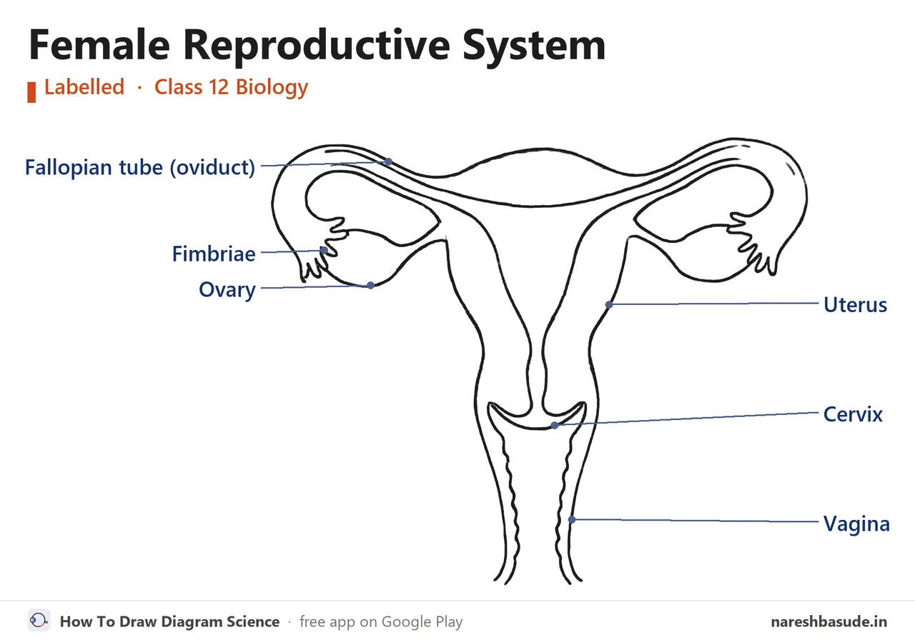
What does the ovary do?
The ovaries are the primary sex organs of the female. They do two jobs: they make the female gamete (ovum), and they make several steroid hormones called ovarian hormones.
There is one ovary on each side of the lower belly. Each ovary is about 2 to 4 cm long. It is joined to the pelvic wall and the uterus by ligaments (strong cords of tissue).
A thin layer called the epithelium covers the ovary. The filling inside the ovary is called the stroma, like the pulp inside a fruit.
The stroma has two parts: the cortex on the outside, and the medulla in the middle. Follicles (the egg's nests, Part 8) at different stages sit inside this stroma.
What are the parts of the fallopian tube?
The fallopian tubes, the uterus and the vagina together are called the female accessory ducts. Each fallopian tube is about 10 to 12 cm long, and it goes from near the ovary to the uterus.
Walk from the ovary towards the uterus, and you meet them in this order:
- Fimbriae: the fingers on the edge of the mouth. They catch the egg that leaves the ovary and take it inside.
- Infundibulum: the wide, funnel-like mouth of the tube, with the fimbriae on its edge
- Ampulla: the wide part of the tube. Fertilisation happens here (Part 10).
- Isthmus: the last, thin part, which joins the uterus
NCERT counts only 3 parts of the tube: the infundibulum, the ampulla and the isthmus. The fimbriae are not a separate part; they are the fingers on the edge of the infundibulum.
Trick: the order from ovary to uterus is "Fresh Idli Aur Imli". Fimbriae, Infundibulum, Ampulla, Isthmus.
RiyaSir, what is the work of the fimbriae? The egg will go into the tube by itself, right?
SirNo, Riya. The tube is not stuck to the ovary, so an egg could fall into the empty space of the belly. The fimbriae catch it and take it into the tube, like a fielder catching a ball.
Don't mix them up: fertilisation happens in the ampulla, not in the uterus. Implantation happens later in the uterus.
Hindi medium? See the Hindi names (tap to open)
| English | Hindi name | Meaning of the pieces |
|---|---|---|
| Ovary | अंडाशय | अंड (egg) + आशय (place to keep) |
| Fallopian tube / oviduct | अंडवाहिनी | अंड + वाहिनी (carrying tube) |
| Fimbriae | झालर | A fringe on the edge, the fingers |
| Infundibulum | कीपक | कीप (funnel) |
| Ampulla | तुंबिका | The wide part |
| Isthmus | संकीर्ण पथ | संकीर्ण (narrow) + पथ (path) |
| Stroma | पीठिका | The inner filling of the ovary |
Revise: 6 cards (tap one to flip it)
Ovary
Primary female sex organ: ovum + ovarian hormones. 2-4 cm.
Stroma
The inner part of the ovary: cortex outside, medulla inside.
Fallopian tube
10-12 cm. Infundibulum (fimbriae on its edge), ampulla, isthmus.
Fimbriae
Finger-like ends. They catch the egg.
Ampulla
The wide part of the tube. Fertilisation happens here.
Accessory ducts
Fallopian tubes, uterus, vagina.
Check yourself
Who catches the egg after ovulation?
The fimbriae, the fingers at the end of the tube.
In which part of the fallopian tube does fertilisation happen?
The ampulla, the wide part of the tube.
Which part of the fallopian tube joins the uterus?
The isthmus, the last thin part of the tube.
Now the baby's home: the uterus. Its wall has 3 layers, and one of them changes every month.
Which layer, and why? In the next part.
5What is the uterus (garbhashaya) like?
In short: the uterus is the place where the baby grows for 9 months. Its wall has 3 layers: a thin cover outside, thick muscle in the middle, and a soft lining inside that changes every month. The uterus opens into the vagina below through the cervix.
Guess firstWhat is the shape of the uterus like?
An upside-down pear! It is wide on top and narrow below. The narrow part at the bottom is the cervix.
Where is the uterus, and what does it look like?
There is only one uterus. It is also called the womb (garbhashaya). It is shaped like an upside-down pear, and ligaments join it to the pelvic wall.
The uterus opens into the vagina through a narrow part at the bottom. This narrow part is called the cervix. The channel inside the cervix is called the cervical canal.
The cervical canal and the vagina together make the path through which the baby is born. This is called the birth canal.
What are the 3 layers of the uterus wall?
| Layer | Where | What it is like and what it does |
|---|---|---|
| Perimetrium | Outermost | A thin, membrane-like layer |
| Myometrium | In the middle | Thick muscle (smooth muscle). It squeezes hard at the time of birth. |
| Endometrium | Innermost | A soft layer with glands. It changes every month in the menstrual cycle. |
Trick: Peri = outside (like perimeter). Myo = muscle. Endo = inside.
Think of the endometrium as a soft bed laid out for the baby (the uterus's soft lining). Every month it becomes thick. If no baby comes, it breaks down and flows out.
This is the period (menstruation). Its full story is in Part 9.
The outer parts (external genitalia)
The outer reproductive parts of the female are called the external genitalia. For the exam, remember each name with one line:
| Name | What it is |
|---|---|
| Mons pubis | A cushion of fat, covered with skin and hair |
| Labia majora | Large fleshy folds that come down from the mons pubis and surround the opening of the vagina |
| Labia minora | A pair of smaller folds below the labia majora |
| Hymen | A thin membrane that sometimes partly covers the opening of the vagina |
| Clitoris | A small finger-like part, at the place where the two labia minora join at the top, above the urinary opening |
What is the truth about the hymen?
In society there is a wrong belief about the hymen. NCERT says clearly: whether the hymen is present or not is not a sure proof of virginity or sexual experience.
The hymen often tears the first time of coitus (sexual intercourse). But it can also tear from a fall, a jerk, using a tampon, or sports like cycling and horse riding. In some women it stays even after coitus.
Exam True/False: "The presence or absence of the hymen is not a reliable proof of virginity." This statement is True (NCERT exercise 16g).
RiyaSir, the cervix and the vagina are the same thing, right?
SirNo. The cervix is the narrow lower part of the uterus. The vagina is the tube below it, which opens outside.
SirThe canal of the cervix and the vagina together make the birth canal.
Don't mix them up: at birth, the myometrium squeezes. Every month, the endometrium changes. Do not write them the wrong way round.
Hindi medium? See the Hindi names (tap to open)
| English | Hindi name | Meaning of the pieces |
|---|---|---|
| Uterus | गर्भाशय | गर्भ (baby) + आशय (place to keep) |
| Cervix | गर्भाशय ग्रीवा | गर्भाशय + ग्रीवा (neck) |
| Vagina | योनि | The tube from the uterus to the outside |
| Birth canal | जन्म नाल | जन्म (birth) + नाल (path) |
| Perimetrium | परिगर्भाशय | परि (outside) + गर्भाशय |
| Myometrium | गर्भाशय पेशी स्तर | The layer of पेशी (muscle) |
| Endometrium | गर्भाशय अंतःस्तर | The layer of अंतः (inside) |
| Hymen | योनिच्छद | योनि + छद (covering membrane) |
Revise: 6 cards (tap one to flip it)
Uterus
The womb. Shaped like an upside-down pear. The baby grows here.
Cervix
The narrow lower part of the uterus. Opens into the vagina.
Birth canal
Cervical canal + vagina.
Myometrium
The thick middle muscle. Squeezes at birth.
Endometrium
The inner layer with glands. Changes every month.
Hymen
A thin membrane at the opening of the vagina. Not a sure proof of virginity.
Check yourself
Which layer of the uterus changes every month in the menstrual cycle?
The endometrium, the innermost layer.
Which two parts make the birth canal?
Cervical canal + vagina.
Which layer squeezes hard at the time of birth?
The myometrium, the thick middle layer of muscle.
We have seen the home and the paths. Now the real question: how is a sperm made from one cell inside the testis?
It uses the same meiosis you saw in Chapter 1, only the names are new. In the next part.
6How are sperm made (spermatogenesis)?
In short: germ cells in the tubule wall first increase their number by mitosis. Then some of them do meiosis: 1 cell becomes 4 cells, and chromosomes go from 46 to 23. These 4 round cells later become sperm with tails.
Guess firstAfter meiosis, how many sperm does one germ cell make?
4 sperm, and all four are alike. For the egg the count is different. You will see that in Part 8.
What is gametogenesis?
The testis and the ovary are called primary sex organs, because they make the gametes. The testis makes sperm and the ovary makes the ovum. Making gametes is called gametogenesis.
Making sperm in the testis is called spermatogenesis. It starts at puberty.
The 5 stations of sperm making
Think of a factory line. One cell passes through 5 stations on the line. At each station its name changes.
- Spermatogonia (46): germ cells in the tubule wall. They increase their number by mitosis.
- Primary spermatocyte (46): the same spermatogonium, with a new name as soon as it starts meiosis.
- Secondary spermatocytes (23): 2 alike cells made by the first division of meiosis (meiosis I).
- Spermatids (23): 4 alike cells made by the second division of meiosis (meiosis II).
- Spermatozoa, or sperm (23): the spermatid changes shape and gets a tail.
Not all spermatogonia move ahead. Only a few become primary spermatocytes, and the rest stay as a stock. That is why a man keeps making sperm till old age.
The secret in the names: "-gonia" = the first cells, "-cyte" = cell, "-tid" = almost ready, "-zoa" = animal, something alive that moves.
When does 46 become 23?
A spermatogonium is diploid: 46 chromosomes (2n). The first division of meiosis (meiosis I) cuts the chromosomes to half. That is why it is called reduction division.
So secondary spermatocytes have only 23 chromosomes. They are haploid (n).
Meiosis II only makes 2 cells into 4. Spermatids also keep 23.
| Cell | Chromosomes | Type |
|---|---|---|
| Spermatogonium | 46 | Diploid (2n) |
| Primary spermatocyte | 46 | Diploid (2n) |
| Secondary spermatocyte | 23 | Haploid (n) |
| Spermatid | 23 | Haploid (n) |
| Sperm | 23 | Haploid (n) |
NCERT question: how many chromosomes do spermatids have? Answer: 23.
How does a round cell become a sperm with a tail?
A spermatid is round, and it cannot swim. Its shape changes, and it gets a head and a tail. This change is called spermiogenesis.
After they form, the heads of the sperm stay buried in the Sertoli cells. Then the sperm are released into the tubule. This release is called spermiation.
Trick: spermiogenesis = forming (getting a new shape). spermiation = freedom (getting free from the Sertoli cell).
RiyaSir, spermatogenesis and spermiogenesis are the same, right? Even the spelling is almost the same.
SirNo! Spermatogenesis is the whole journey, from spermatogonia to sperm. Spermiogenesis is only the last station: a spermatid changes shape to become a sperm.
NCERT exercise 8: spermiogenesis = spermatids turning into sperm. Spermiation = release of sperm from the seminiferous tubules.
Why does all this start only at puberty?
The reason is hormones. A hormone is a chemical message of the body. It travels in the blood from one place to another.
At puberty, a part of the brain called the hypothalamus starts releasing more of one hormone. Its name is GnRH.
The full name of GnRH is gonadotropin releasing hormone.
GnRH goes to a small gland under the brain, called the anterior pituitary.
The pituitary then releases 2 hormones: LH (luteinising hormone) and FSH (follicle stimulating hormone).
Together, LH and FSH are called gonadotropins.
- Hypothalamus: GnRH
- Anterior pituitary: LH and FSH
- LH → Leydig cells → make androgens → androgens boost spermatogenesis
- FSH → Sertoli cells → release some special chemicals (factors) that help spermiogenesis
Trick: LH → Leydig. FSH → Sertoli. In each pair the first letter matches.
NCERT exercise 7: hormones that control spermatogenesis: GnRH, LH, FSH and androgens.
Hindi medium? See the Hindi names (tap to open)
| English | Hindi name | Meaning of the pieces |
|---|---|---|
| Spermatogenesis | शुक्राणुजनन | शुक्राणु (sperm) + जनन (making) |
| Spermatogonia | शुक्राणुजन | The first cells of sperm |
| Primary spermatocyte | प्राथमिक शुक्राणुकोशिका | प्राथमिक (first) + शुक्राणु + कोशिका (cell) |
| Secondary spermatocyte | द्वितीयक शुक्राणुकोशिका | द्वितीयक (second) + शुक्राणुकोशिका |
| Spermatid | शुक्राणुप्रसू | Almost ready sperm |
| Spermiogenesis | स्पर्मियोजेनेसिस | Write the English name: a spermatid becoming a sperm |
| Spermiation | स्पर्मिएशन | Write the English name: release of sperm |
| Hypothalamus | हाइपोथैलेमस | A part of the brain |
| Pituitary | पीयूष ग्रंथि | The small gland under the brain |
Revise: 8 cards (tap one to flip it)
Spermatogonia
Germ cells (46). They increase their number by mitosis.
Primary spermatocyte
46. Does meiosis I.
Secondary spermatocyte
23 (haploid). Does meiosis II.
Spermatid
23. Round, not yet a sperm.
Spermiogenesis
A spermatid becomes a sperm (head + tail).
Spermiation
The sperm get free from the Sertoli cells and enter the tubule.
GnRH
Hormone of the hypothalamus. It signals the pituitary to release LH and FSH.
LH / FSH
LH → Leydig → androgens. FSH → Sertoli → helps spermiogenesis.
Check yourself
How many chromosomes does a secondary spermatocyte have?
23. Meiosis I (reduction division) cut the chromosomes to half.
What is the change of a spermatid into a sperm called?
Spermiogenesis. Spermiation is the release, and spermatogenesis is the whole journey.
How many spermatids come from one primary spermatocyte?
4. Meiosis I gives 2, and then meiosis II gives 4.
LH acts on which cells?
Leydig cells. LH → Leydig → androgens. FSH acts on the Sertoli cells.
The sperm is made. But what does it look like? Its head, middle part and tail each have a different job.
And where does it get the power to swim? In the next part.
7What does a sperm look like, and what is semen?
In short: a sperm has 4 parts: head, neck, middle piece and tail. The head carries the information (nucleus), the middle has the energy engine, and the tail does the swimming. Fluid from glands on the way mixes with sperm to make semen.
Guess firstAbout how many sperm come out at one time?
200 to 300 million! But only one of them reaches the egg and joins it. See below why so many are needed.
What are the 4 parts of a sperm?
A sperm is so small that you can see it only with a microscope. Think of it as a tiny swimming rocket. A thin skin (plasma membrane) covers the whole sperm.
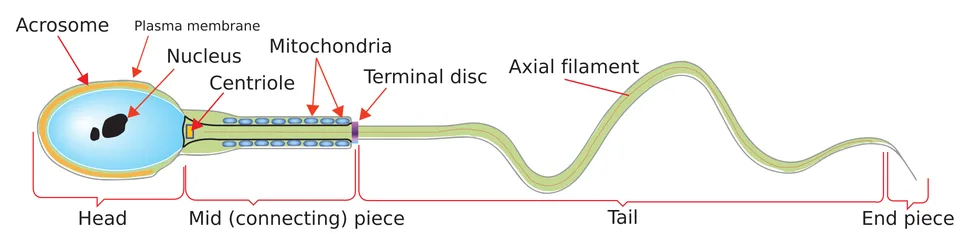
| Part | What is in it | Job |
|---|---|---|
| Head | A long nucleus (23 chromosomes), and a cap in front: the acrosome | The nucleus carries the father's information. The acrosome's enzymes help the sperm enter the egg. |
| Neck | The joint between the head and the middle piece | It joins the two |
| Middle piece | Many mitochondria | They make the energy to move the tail |
| Tail | A long tail | It lashes to and fro and pushes the sperm ahead |
Mitochondria are called the power house of the cell. They make energy from food. A sperm must swim (motility) to meet the egg, so the middle piece has many mitochondria.
Trick: head = information + key (nucleus + acrosome). Middle = battery (mitochondria). Tail = oar (for swimming).
Why so many sperm?
About 200 to 300 million sperm come out at one time. For normal fertility, two conditions must be met:
- At least 60% of the sperm have a normal shape and size.
- At least 40% of the sperm swim fast (vigorous motility).
What do the glands on the way add?
Three kinds of glands add their fluid on the sperm's path. They are called male accessory glands.
| Gland | How many |
|---|---|
| Seminal vesicles | 2 (one pair) |
| Prostate | 1 |
| Bulbourethral glands | 2 (one pair) |
Trick: the count is "2-1-2": seminal vesicles 2, prostate 1, bulbourethral 2.
The fluid of these glands together is called seminal plasma. It has 3 special things: fructose (a sugar, the sperm's fuel), calcium, and some enzymes. The fluid of the bulbourethral glands also keeps the penis slippery (lubricated).
Seminal plasma and sperm together are called semen.
What do the ducts and glands do?
Not only the glands: the walls of the ducts (epididymis and vas deferens) also add a little fluid. The fluid of the epididymis, vas deferens, seminal vesicle and prostate helps the sperm mature and swim.
The ducts also store the sperm and carry them outside.
Androgens (the hormones of the Leydig cells) keep all these ducts and glands working well.
NCERT exercise 11: the ducts store and transport the sperm. The glands make seminal plasma, which matures the sperm, helps them swim, and gives fuel (fructose).
RiyaSir, semen and sperm are the same thing, right?
SirNo! A sperm is a single cell. Semen is the whole liquid: crores of sperm + the fluid of the glands (seminal plasma).
Don't mix them up: the mitochondria of the middle piece make the energy, not the tail. The tail only moves with that energy.
Hindi medium? See the Hindi names (tap to open)
| English | Hindi name | Meaning of the pieces |
|---|---|---|
| Acrosome | अग्रपिंडक | अग्र (front) + पिंडक (cap-like part) |
| Middle piece | मध्य भाग | The middle part |
| Mitochondria | सूत्रकणिका | The power house of the cell |
| Seminal vesicle | शुक्राशय | शुक्र (semen) + आशय (place to keep) |
| Prostate | पुरःस्थ ग्रंथि | पुरः (front) + स्थ + ग्रंथि (gland) |
| Semen | वीर्य | Sperm + fluid of the glands |
Revise: 7 cards (tap one to flip it)
Head
Nucleus (23) + acrosome (enzymes, to enter the egg).
Middle piece
Mitochondria: energy for the tail.
Tail
Makes the sperm swim (motility).
Normal fertility
200-300 million sperm; 60% normal shape, 40% fast swimmers.
Accessory glands
Seminal vesicles (2), prostate (1), bulbourethral (2).
Seminal plasma
Fructose, calcium, enzymes.
Semen
Sperm + seminal plasma.
Check yourself
What does the acrosome do?
The enzymes of the acrosome help the sperm enter the egg. The middle piece gives energy, and the nucleus carries the information.
For normal fertility, at least what % of sperm must swim fast?
40% fast swimmers. The 60% condition is for normal shape and size.
Which of these is NOT in seminal plasma?
Sperm. Seminal plasma is only the fluid of the glands. Sperm + seminal plasma = semen.
Now the most surprising fact on the mother's side: the making of eggs starts before the girl is even born!
And unlike sperm, 1 cell does not make 4 eggs. How many does it make? In the next part.
8How is the egg (ovum) made (oogenesis)?
In short: the making of the egg starts before the girl herself is born, and then it pauses. Each egg lives in a small nest (follicle) that slowly grows bigger. In the end the big nest bursts and the egg comes out: this is called ovulation.
Guess firstWhen do the cells that make eggs form in a girl's ovary?
Before birth! After birth no new egg-mother cell forms. This is a very different story from sperm.
When does the egg's story begin?
Making a mature egg in the ovary is called oogenesis. It is very different from spermatogenesis.
While the girl is still in her mother's womb (as a foetus), about 20 lakh egg-mother cells form in each ovary. They are called oogonia. After birth no new oogonium forms.
These oogonia start meiosis, but they stop at the first stage of the first division (meiosis I), called prophase-I. A cell stopped like this is called a primary oocyte.
They stay paused for years. After puberty, when their nest grows, they move ahead.
The egg and its nest: two lines together
Think of two lines. One line is the egg. The other line is its nest (follicle), a layer of cells around the egg that grows with it.
- Primary follicle: one layer of granulosa cells around the primary oocyte
- Secondary follicle: more layers of granulosa, and a new layer outside, the theca
- Tertiary follicle: a space full of fluid forms inside, called the antrum. The theca becomes two layers: theca interna inside, theca externa outside.
- Graafian follicle: the biggest, mature follicle
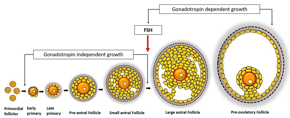
Don't mix them up: "primary" and "secondary" are used in two places: for the egg cell (oocyte) and for the follicle. See both together in the table below.
| Follicle (nest) | Egg cell inside |
|---|---|
| Primary follicle | Primary oocyte (paused at meiosis I) |
| Secondary follicle | Primary oocyte (still paused) |
| Tertiary follicle | The primary oocyte grows, finishes meiosis I → secondary oocyte + first polar body |
| Graafian follicle | Secondary oocyte, which makes a zona pellucida around itself |
From birth to puberty, many follicles are lost. At puberty only 60,000 to 80,000 primary follicles are left in each ovary.
Why is the egg's division not equal?
Inside the tertiary follicle the primary oocyte grows big. It finishes its first division (meiosis I). But this division is not equal.
One big cell forms: the secondary oocyte (haploid). And one very small cell forms: the first polar body. Almost all the food-rich part (cytoplasm) goes to the secondary oocyte.
Think of sharing a roti: the whole roti goes to one, and only a crumb to the other. The benefit: the future baby (zygote) gets food at the start.
Does the first polar body divide further or die? NCERT says: this is not sure yet.
What is ovulation?
Now the secondary oocyte makes a new covering around itself, called the zona pellucida. The follicle has now become a Graafian follicle.
The Graafian follicle bursts, and the secondary oocyte (ovum) comes out of the ovary. This is called ovulation.
Remember: what comes out in ovulation is the secondary oocyte. Meiosis II is finished only when a sperm arrives and meets it (Part 10).
Sperm making and egg making: the difference
| Point | Spermatogenesis | Oogenesis |
|---|---|---|
| When it starts | At puberty | Before birth (in the foetus) |
| From one mother cell | 4 sperm | 1 ovum (the rest are small polar bodies) |
| Division | Equal | Not equal (big oocyte, small polar body) |
| Till when | Till old age | Till about 50 years |
RiyaSir, is a polar body also an egg? Can it also make a baby?
SirNo. A polar body has chromosomes, but almost no food (cytoplasm). It cannot make a baby.
Don't mix them up: the follicle and the oocyte are different. The oocyte is the egg. The follicle is the nest of cells around it.
Hindi medium? See the Hindi names (tap to open)
| English | Hindi name | Meaning of the pieces |
|---|---|---|
| Oogenesis | अंडजनन | अंड (egg) + जनन (making) |
| Oogonia | अंडजननी | The first cells of the egg |
| Primary oocyte | प्राथमिक अंडक | प्राथमिक (first) + अंडक |
| Secondary oocyte | द्वितीयक अंडक | द्वितीयक (second) + अंडक |
| Polar body | ध्रुवीय पिंड | ध्रुव (end) + पिंड (small ball) |
| Follicle | पुटक | The nest of the egg |
| Graafian follicle | ग्राफी पुटक | The mature, biggest follicle |
| Ovulation | अंडोत्सर्ग | अंड + उत्सर्ग (release) |
Revise: 8 cards (tap one to flip it)
Oogonia
Egg-mother cells. They form before birth, and no new ones form later.
Primary oocyte
A cell paused at the start of meiosis I.
Primary follicle
Primary oocyte + one layer of granulosa cells.
Tertiary follicle
Antrum (fluid space) + theca interna and externa.
Secondary oocyte
Big haploid cell after meiosis I. A small first polar body comes with it.
Graafian follicle
The biggest, mature follicle. It bursts and releases the egg.
Zona pellucida
The new covering around the secondary oocyte.
Follicles at puberty
60,000-80,000 primary follicles in each ovary.
Check yourself
The antrum (fluid space) marks which follicle?
The tertiary follicle. Theca interna and externa also form in it.
What comes out of the ovary at ovulation?
The secondary oocyte. Meiosis II is finished when a sperm meets it.
About how many primary follicles are left in each ovary at puberty?
60,000 to 80,000. At the start there were about 20 lakh oogonia, and the rest were lost on the way.
Ovulation happens once a month. But who tells the ovary when? And why does the lining of the uterus change every month?
A 28-day clock runs all of this: the menstrual cycle. In the next part.
9What happens every month in the menstrual cycle?
In short: about every 28 days the ovary releases an egg, and the uterus's soft bed (lining), the endometrium, gets ready for a baby. If no baby forms, the lining breaks down and flows out: these are periods. A clock of hormones runs all of this.
Guess firstIn a 28-day cycle, around which day is the egg released (ovulation)?
In the middle, around day 14. See the whole clock below.
What is the menstrual cycle?
In female primates (monkeys, apes and humans) the reproductive cycle repeats every month. It is called the menstrual cycle. The days from one period to the next make one cycle: on average about 28-29 days.
The first period comes at puberty: it is called menarche. Around the age of 50 the cycle stops: this is called menopause.
Periods are a normal and healthy process of the body, not a matter of shame. Regular periods from menarche to menopause show that the reproductive system is working well.
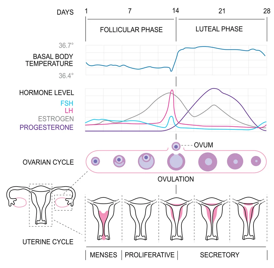
What are the 4 phases of the cycle?
First, the menstrual phase (day 1 to about day 5): the lining of the endometrium and its blood vessels break down. It flows out through the vagina as a liquid. This lasts 3 to 5 days.
Second, the follicular phase: in the ovary the primary follicles grow into a Graafian follicle. At the same time the endometrium in the uterus starts to thicken again. This thickening is called proliferation.
In this phase the pituitary hormones, LH and FSH, rise slowly. They make the follicles grow, and the growing follicles make a hormone called estrogen. Estrogen helps to thicken the uterus lining (endometrium) again.
Third, the ovulatory phase (around day 14): LH and FSH both rise to their peak, but LH rises the most and very suddenly. This sudden jump of LH is called the LH surge. The LH surge bursts the Graafian follicle, and the egg comes out (ovulation).
Fourth, the luteal phase: the part of the burst Graafian follicle that is left turns into a yellow spot, called the corpus luteum. It makes a lot of progesterone.
Progesterone keeps the endometrium (the lining) in good shape. Such a lining is needed for the baby to attach (implantation) in the uterus and for pregnancy.
What if a baby forms, and what if it does not?
- The egg was not fertilised: the corpus luteum breaks down by the end of the luteal phase. Progesterone falls, the lining breaks down, periods come, and a new cycle starts.
- Pregnancy happened: all the events of the cycle stop. Periods do not come during pregnancy.
So a missed period can be a sign of pregnancy. But it can also come from other reasons, such as stress or poor health.
NCERT exercise 17: the menstrual cycle is controlled by the pituitary hormones (LH, FSH) and the ovary hormones (estrogen, progesterone). Exercise 16(f): "The menstrual cycle stops during pregnancy" = True.
RiyaSir, where does the blood of periods come from? Is it an injury?
SirNo, it is not an injury. The inner layer of the uterus (endometrium) and its tiny blood vessels break down and come out every month, because no baby formed. A new layer forms the next month.
Don't mix them up: the corpus luteum makes progesterone, not the follicle. Growing follicles make estrogen.
Menstrual hygiene (NCERT's advice):
- Bathe daily and keep yourself clean.
- Use a sanitary napkin, or a clean pad made at home.
- Change the napkin or pad every 4 to 5 hours, as needed.
- Wrap the used napkin in old paper and throw it away properly. Do not throw it in the toilet or in the open.
- Wash your hands with soap after touching the napkin.
Hindi medium? See the Hindi names (tap to open)
| English | Hindi name | Meaning of the pieces |
|---|---|---|
| Menstrual cycle | आर्तव चक्र | आर्तव (monthly flow) + चक्र (cycle) |
| Menarche | रजोदर्शन | The first period |
| Menopause | रजोनिवृत्ति | The stopping of periods |
| Corpus luteum | पीत पिंड | पीत (yellow) + पिंड (spot) |
| Ovulation | अंडोत्सर्ग | अंड + उत्सर्ग (release) |
| Progesterone | प्रोजेस्टेरोन | Just the English name |
Revise: 7 cards (tap one to flip it)
Menarche / menopause
The first period (puberty) / the stopping of periods (about 50 years).
Menstrual phase
3-5 days. The endometrium and blood vessels break down and flow out.
Follicular phase
The follicle grows, the endometrium thickens, estrogen rises.
LH surge
The sudden jump of LH around day 14. It causes ovulation.
Corpus luteum
The left-over part of the burst follicle. Makes progesterone.
Progesterone
Keeps the endometrium in shape, needed for pregnancy.
No baby formed
Corpus luteum fades, endometrium breaks down, periods.
Check yourself
The sudden rise of which hormone causes ovulation?
The LH surge, around day 14.
Which hormone does the corpus luteum make?
Progesterone, which keeps the endometrium in good shape.
When do periods come?
When there is no fertilisation: the corpus luteum fades and the endometrium breaks down and flows out.
Now the biggest moment of the story: the egg is in the fallopian tube, and crores of sperm are swimming towards it.
Only one gets in. How are the others stopped? And who decides if the baby is a boy or a girl?
Find out in the next part.
10Fertilisation: how do the sperm and egg meet?
In short: sperm swim up to the ampulla of the fallopian tube, where the egg is waiting. Only one sperm gets inside the egg, and the door shuts at once for the rest. The two cells then join into one.
Guess firstWhether the baby is a boy or a girl is decided by whose cell?
The father's sperm! Scientifically, blaming the mother for a daughter is wrong. See why below.
How do the sperm reach the egg?
During coitus (sexual intercourse), semen is released into the vagina. This is called insemination.
Sperm swim fast. They pass through the cervix into the uterus, and finally reach the ampulla part of the fallopian tube.
The egg released from the ovary also travels through the tube to the ampulla. Fertilisation happens right here.
Remember: fertilisation happens only if the sperm and the egg are in the ampulla at the same time. That is why not every coitus leads to pregnancy.
Why does only one sperm go in?
There is a thin layer around the egg: the zona pellucida (Part 8). The moment one sperm touches this layer, the layer changes. This change blocks the way for the other sperm.
Think of it like a one-time password: once used, it is of no use again. That is why one egg is fertilised by only one sperm.
The cap on the sperm's head, the acrosome (Part 7), releases its enzymes. These help the sperm cut through the zona pellucida and the egg's outer layer.
The egg's half-finished division is now completed
Remember Part 8? At ovulation, a secondary oocyte came out, and its second division (meiosis II) was half done. The moment a sperm enters, this division is completed.
This division is also unequal. A small second polar body forms, and a big haploid ovum. This ripe, ready ovum is also called the ootid.
Now the sperm's nucleus (23) and the ovum's nucleus (23) join. The result is a zygote with 46 chromosomes.
Because it has the full 46, the zygote is diploid.
NCERT question: how many chromosomes does a zygote have? Answer: 46.
Boy or girl: who decides?
Two of the 46 chromosomes are special, and they are called sex chromosomes. A woman has XX, and a man has XY.
- All of the mother's eggs carry one X. The mother has only X to give.
- Half (50%) of the father's sperm carry X, and half (50%) carry Y.
| In the egg | In the sperm | Zygote | Baby |
|---|---|---|---|
| X | X | XX | Girl |
| X | Y | XY | Boy |
So whether the baby is a boy or a girl is decided at this very moment, and it depends on the father's sperm. Scientifically, the father decides the baby's sex, not the mother.
RiyaSir, then why do people blame the mother when a daughter is born?
SirBecause they do not know this science. The mother has only X to give, and Y is found only in the father's sperm.
SirSo blaming the mother is completely wrong. This is also NCERT exercise 19.
How many eggs in a month? How do twins form?
A human ovary usually releases 1 egg every month.
- Identical twins (exactly alike): 1 egg, 1 sperm, 1 zygote. Later the early embryo splits into 2.
- Fraternal twins (look different): 2 eggs were released, and each met a different sperm.
NCERT exercise 20-21: identical twins = 1 egg, fraternal twins = 2 eggs. If a female dog had 6 puppies, about 6 eggs were released, because each puppy came from a separate egg.
Don't mix them up: fertilisation happens in the ampulla of the fallopian tube, not in the uterus.
Hindi medium? See the Hindi names (tap to open)
| English | Hindi name | Meaning of the pieces |
|---|---|---|
| Insemination | वीर्यसेचन | वीर्य (semen) + सेचन (to put in) |
| Fertilisation | निषेचन | The meeting of the sperm and the egg |
| Zona pellucida | पारदर्शी अंडावरण | पारदर्शी (see-through) + आवरण (covering) |
| Polar body | ध्रुवीय पिंड | ध्रुव (end) + पिंड (body) |
| Zygote | युग्मनज | युग्म (pair) + ज (made from) |
| Sex chromosomes | लिंग गुणसूत्र | लिंग (sex) + गुणसूत्र (chromosome) |
Revise: 6 cards (tap one to flip it)
Insemination
Semen being released into the vagina.
Where do they meet?
The ampulla of the fallopian tube. Only if both arrive together.
Zona pellucida
Closes for the others as soon as the first sperm touches it.
When is meiosis II completed?
As soon as the sperm enters. Second polar body + ovum (ootid).
Zygote
23 + 23 = 46 chromosomes, diploid.
Boy or girl
X sperm = XX girl. Y sperm = XY boy. The father decides.
Check yourself
Why does an egg not get fertilised by more than one sperm?
The zona pellucida changes as soon as the first sperm touches it, and this blocks the others.
What will an XY zygote become?
A boy. The Y came from the father's sperm.
How many eggs are needed for identical twins?
1 egg. Its early embryo splits into 2 later. Fraternal twins need 2 eggs.
The zygote is made, but it is still in the fallopian tube. The way to the uterus takes a few days, and on the way it divides again and again until it looks like a ball.
How does it then attach to the wall of the uterus? In the next part.
11How does the zygote attach to the uterus?
In short: the zygote moves towards the uterus and divides into a ball of cells. In the uterus it becomes a hollow ball (the blastocyst) that sinks into the soft bed (the endometrium). This is implantation, and pregnancy starts here.
Guess firstThe ball of 8-16 cells made by the zygote's divisions is named after a fruit. Which one?
Mulberry! The ball is called a morula, which is the Latin name for mulberry. It even looks like one.
What does the zygote do on the way?
The zygote moves towards the uterus through the isthmus part of the fallopian tube. As it moves, it keeps dividing by mitosis: 2, then 4, then 8, then 16 cells.
This dividing is called cleavage. The small cells that form are called blastomeres.
When there are 8 to 16 blastomeres, the ball looks like a mulberry. This stage is called the morula.
What is a blastocyst?
The morula keeps dividing. By the time it reaches the uterus, it has become a hollow ball. This is the blastocyst.
Its cells are in two groups:
- Trophoblast: the outer layer. This is the part that sticks to the wall of the uterus.
- Inner cell mass: a clump of cells inside, stuck to the trophoblast. This later becomes the baby (embryo).
Trick: "tropho" means food. The trophoblast later helps to make the placenta, which brings food to the baby. Inner cell mass = the real player inside: the baby.
How does implantation happen?
The trophoblast sticks to the uterus's soft bed, the endometrium. Then the endometrium cells divide fast and cover the blastocyst, like soil covers a seed.
Now the blastocyst is buried inside the endometrium. This is called implantation. Pregnancy starts with implantation.
- Zygote (in the fallopian tube)
- Cleavage: 2, 4, 8, 16 blastomeres
- Morula (8-16 cells)
- Blastocyst: trophoblast + inner cell mass
- Implantation: sinking into the endometrium
RiyaSir, the zygote was one cell. Does the baby get bigger as the cells divide?
SirNot at first! In cleavage the number of cells grows, but each new cell is smaller. The ball stays about as big as the zygote.
SirReal growth starts after implantation, when food begins to come from the mother.
Don't mix them up: the baby comes from the inner cell mass, not from the trophoblast. The trophoblast helps to stick to the uterus and later to make the placenta (Part 12).
Hindi medium? See the Hindi names (tap to open)
| English | Hindi name | Meaning of the pieces |
|---|---|---|
| Cleavage | विदलन | Dividing |
| Blastomeres | कोरक खंड | The small cells made by dividing |
| Morula | तूतक | तूत (mulberry) like |
| Blastocyst | कोरकपुटी | A hollow ball |
| Trophoblast | पोषकोरक | The outer layer |
| Inner cell mass | अंतर कोशिका समूह | अंतर (inside) + कोशिका समूह (clump of cells) |
| Implantation | अंतर्रोपण | Planting inside |
Revise: 6 cards (tap one to flip it)
Cleavage
The zygote dividing by mitosis while moving in the tube.
Blastomeres
The small cells made by cleavage.
Morula
A mulberry-like ball of 8-16 blastomeres.
Blastocyst
A hollow ball: trophoblast (outside) + inner cell mass (inside).
Inner cell mass
The embryo (baby) forms from this.
Implantation
The blastocyst sinking into the endometrium. Pregnancy starts.
Check yourself
How many blastomeres does a morula have?
8 to 16.
Which part of the blastocyst becomes the baby?
The inner cell mass. The trophoblast sticks to the uterus.
What does the zygote turn into before it implants in the uterus?
The blastocyst. The NCERT fill-in-the-blank asks the same thing.
The baby is now attached inside the mother. But how will it get food and oxygen? It cannot breathe!
For this, a special "connection" forms between the mother and the baby: the placenta. In the next part.
12How does the baby grow during pregnancy?
In short: a placenta forms between the mother and the baby. Through it the baby gets oxygen and food, and its waste goes out. The baby is built slowly over 9 months: first the heart, then the limbs, then everything else.
Guess firstWhich organ of the baby forms first, so that a doctor can hear its beat?
The heart! It forms in just one month. Hearing its beat with a stethoscope is the first sign of the baby.
What is the placenta?
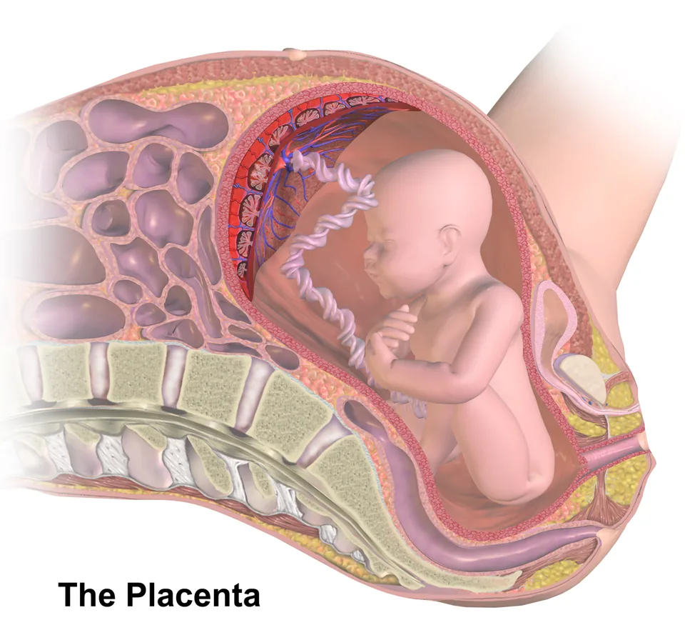
After implantation, small finger-like bumps grow out of the trophoblast. These are called chorionic villi. They are surrounded by uterus tissue and the mother's blood.
The chorionic villi and the uterus tissue lock into each other, like fingers of two hands folded together. This joined part is the placenta.
NCERT's line: the placenta is a "structural and functional unit" between the baby (foetus) and the mother's body, joined in both build and work.
What does the placenta do?
- It brings oxygen and food from the mother's blood to the baby.
- It carries the baby's carbon dioxide and waste back to the mother's blood.
- It joins the baby by a cord, the umbilical cord. Everything goes to and fro through this cord.
- It also makes hormones. A tissue that makes hormones is an endocrine tissue, so the placenta is an endocrine tissue too.
Special hormones of pregnancy
The placenta makes two special hormones: hCG (human chorionic gonadotropin) and hPL (human placental lactogen).
It also makes estrogens and progestogens.
In the last part of pregnancy, the ovary makes one more hormone: relaxin.
Remember: hCG, hPL and relaxin are made only in pregnancy. Trick: "h" = human, and both "h" hormones belong to the placenta. Relaxin alone belongs to the ovary.
In pregnancy, some other hormones in the mother's body also rise many times. This helps the baby grow, brings the needed (metabolic) changes in the mother's body, and keeps the pregnancy safe. For the exam, just remember their names: estrogens, progestogens, cortisol, prolactin, thyroxine.
Where do the baby's body parts come from?
Soon after implantation, the inner cell mass (Part 11) splits into two layers: ectoderm outside and endoderm inside.
Very soon a third layer comes in between: the mesoderm.
These 3 layers later make all the tissues and organs of the grown person.
The inner cell mass has some special cells called stem cells. They have the power to become any tissue or organ of the body.
Trick: Ecto = outside, Endo = inside, Meso = in the middle (think of "medium").
The baby, month by month
A human pregnancy lasts 9 months. Each block of three months is called a trimester.
| When | What forms |
|---|---|
| 1 month | The heart forms. Hearing the beat with a stethoscope is the first sign. |
| End of 2 months | Limbs and fingers (digits) |
| 12 weeks, about 3 months (first trimester) | Most major organ systems are formed. Limbs and the outer genital organs are well formed. |
| 5th month | The baby moves for the first time, and hair appears on the head |
| 24 weeks, about 6 months (second trimester) | Fine hair on the body, the eyelids separate, and eyelashes form |
| 9 months | The baby is fully formed and ready to be born |
NCERT's question: how long are other animals pregnant? Dogs and cats about 2 months, and elephants about 22 months!
RiyaSir, does the mother's blood go straight into the baby?
SirNo, the two bloods do not mix. The mother's blood stays outside the villi, and the baby's blood stays inside. A very thin wall lies between them.
SirOxygen and food pass through this thin wall, like a tea strainer: the tea goes through and the leaves stay behind.
Don't mix them up: relaxin is made by the ovary, not the placenta, and only in the last part of pregnancy.
Hindi medium? See the Hindi names (tap to open)
| English | Hindi name | Meaning of the pieces |
|---|---|---|
| Placenta | अपरा | The link between mother and baby |
| Chorionic villi | जरायु अंकुरक | Finger-like bumps |
| Umbilical cord | नाभि रज्जु | नाभि (navel) + रज्जु (cord) |
| Ectoderm | बहिर्जनस्तर | बहिः (outside) + जनस्तर (the layer that makes) |
| Endoderm | अंतर्जनस्तर | अंतः (inside) + जनस्तर |
| Mesoderm | मध्यजनस्तर | मध्य (middle) + जनस्तर |
| Stem cells | स्टेम कोशिकाएँ | Cells that can become anything |
Revise: 7 cards (tap one to flip it)
Chorionic villi
Finger-like bumps of the trophoblast. With uterus tissue they make the placenta.
Placenta
Oxygen and food in; CO2 and waste out. It also makes hormones.
Umbilical cord
The cord joining the baby and the placenta.
Pregnancy-only hormones
hCG, hPL (placenta), relaxin (ovary).
3 germ layers
Ectoderm (outside), mesoderm (middle), endoderm (inside). All organs come from these.
Stem cells
Cells of the inner cell mass that can make any tissue.
First trimester
12 weeks: most organ systems are formed.
Check yourself
What is the blood-vessel connection (vascular connection) between the baby and the mother?
The placenta. The NCERT fill-in-the-blank asks the same thing.
Which pregnancy hormone does the ovary make?
Relaxin, in the last part of pregnancy. hCG and hPL are made by the placenta.
When does the baby first start to move?
About the 5th month, when hair also appears on the head.
9 months are over. Now how does the baby come out, and who gives the "signal" for birth?
And why is the mother's first milk after birth so special? In the next part.
13How is a baby born, and how is mother's milk made?
In short: after 9 months the baby and the placenta send a "signal". The uterus contracts gently, the mother's pituitary gland releases oxytocin, and the contractions get stronger until the baby is out. By the end of pregnancy, the mother's breasts (mammary glands) start making milk.
Guess firstWhere does the "signal" for birth come from first?
From the baby and the placenta! They tell the body that they are now ready.
How does birth (parturition) happen?
Pregnancy lasts about 9 months. This time is called the gestation period.
At the end, the uterus contracts strongly and the baby comes out. The birth of the baby is called parturition.
The brain and the hormones run it together. "Neuro" means brain and nerves, and "endocrine" means hormones, so it is called a neuroendocrine mechanism.
See the steps:
- The fully formed baby and the placenta send a signal.
- The uterus contracts gently. This is called the foetal ejection reflex.
- This makes the mother's pituitary gland release the hormone oxytocin.
- Oxytocin makes the uterus muscle (myometrium) contract harder.
- Stronger contractions release more oxytocin, and the contractions get stronger still.
- The baby comes out through the birth canal. A little later, the placenta comes out too.
Think of it as a loop: contraction → oxytocin → stronger contraction → more oxytocin. With every round, the force grows.
NCERT question: which injection do doctors give to speed up delivery? Answer: oxytocin.
The NCERT summary says this neuroendocrine mechanism involves cortisol, estrogens and oxytocin. Write all three in exercise 18.
What does a mammary gland (breast) look like inside?
Working mammary glands are the mark of every female mammal (an animal that feeds milk). They come as a pair, and they are one of the female secondary sexual characteristics (changes that come at puberty). They hold milk-making tissue and some fat.
The gland part of each breast is divided into 15 to 20 parts called mammary lobes. Each lobe has small clusters of cells called alveoli. The cells of the alveoli make the milk.
Picture the milk path as a tree. It starts at the grape-like berries (alveoli), then thin twigs, then thick branches, then the trunk, and finally the pipe:
- Alveoli: milk is made and stored here
- Mammary tubules: thin tubes
- Mammary duct: all the tubules of one lobe join up
- Mammary ampulla: several ducts join into a wide space. "Ampulla" means a wide part, which is why the name came up in the fallopian tube too.
- Lactiferous duct
- Nipple: the milk comes out here
Lactation and colostrum
During pregnancy the mammary glands change. By the end, they start making milk. Making milk is called lactation, and it lets the mother feed her newborn baby.
The milk of the first few days is called colostrum. It is full of antibodies: these are things that give the body strength to fight disease. They are very important to protect a newborn.
That is why doctors advise feeding the baby the mother's milk in the first months.
RiyaSir, why does the oxytocin loop never stop by itself?
SirIt keeps growing until the baby is out. The moment the baby is out, the signal stops. That is the special thing about this "positive" loop.
Don't mix them up: oxytocin comes from the mother's pituitary, not from the placenta. And it is the myometrium of the uterus that contracts, not the endometrium.
Hindi medium? See the Hindi names (tap to open)
| English | Hindi name | Meaning of the pieces |
|---|---|---|
| Parturition | प्रसव | The birth of the baby |
| Gestation period | सगर्भता काल | The time of pregnancy |
| Foetal ejection reflex | गर्भ उत्क्षेपण प्रतिवर्त | The signal to push the baby out |
| Mammary gland | स्तन ग्रंथि | स्तन (breast) + ग्रंथि (gland) |
| Alveoli | कूपिकाएँ | Small clusters that make milk |
| Lactation | दुग्धस्रवण | दुग्ध (milk) + स्रवण (flowing out) |
| Colostrum | खीस | The milk of the first days |
Revise: 7 cards (tap one to flip it)
Parturition
The birth of the baby, by strong contractions of the uterus.
Foetal ejection reflex
The gentle contraction of the uterus, set off by the signal from the baby and placenta.
Oxytocin
From the mother's pituitary. Makes the uterus contract hard. Also given as an injection for delivery.
Mammary lobes
15-20 in each breast. They hold the alveoli.
The milk path
Alveoli, tubules, duct, ampulla, lactiferous duct, nipple.
Lactation
Making milk, from the end of pregnancy.
Colostrum
The first days' milk. Full of antibodies.
Check yourself
Which hormone makes the uterus contract hard during delivery?
Oxytocin, from the mother's pituitary.
Why is colostrum special?
Antibodies! They give a newborn the strength to fight disease.
Who makes the milk in a mammary gland?
The cells of the alveoli. The other tubes only carry the milk onwards.
The whole story is done: from two cells to a new person, and up to the first feed.
Now use the quick revision below to lock it all in.
14Quick revision: the night before the exam
In short: the whole chapter on one screen. First the story in one line, then the hormones, the numbers, the NCERT fill-in-the-blanks, and the common mistakes.
The whole story in one line
- Testis: spermatogonia (46) → meiosis → 4 sperm (23)
- Ovary: oogonia (in the foetus) → primary oocyte (stopped at prophase-I) → secondary oocyte (ovulation, day 14)
- Insemination → sperm reach the ampulla
- Fertilisation (ampulla): zygote (46), boy or girl decided here
- Cleavage → morula → blastocyst → implantation (endometrium)
- Placenta → 9 months of gestation
- Oxytocin → parturition → lactation (colostrum)
Hormones: which, from where, what job?
| Hormone | From where | Job |
|---|---|---|
| GnRH (gonadotropin releasing hormone) | Hypothalamus | Tells the pituitary to release LH and FSH |
| LH (luteinising hormone) | Anterior pituitary | Male: Leydig → androgens. Female: LH surge → ovulation |
| FSH (follicle stimulating hormone) | Anterior pituitary | Male: Sertoli → spermiogenesis. Female: grows the follicle |
| Androgens | Leydig cells | Run spermatogenesis, the ducts and the glands |
| Estrogen | Growing follicles | Thickens the endometrium again |
| Progesterone | Corpus luteum | Keeps the endometrium in place |
| hCG, hPL | Placenta | Only in pregnancy |
| Relaxin | Ovary (end of pregnancy) | Only in pregnancy |
| Oxytocin | Mother's pituitary | Makes the uterus contract at birth |
NCERT exercise 1: fill in the blanks
- Humans reproduce: sexually. Humans are: viviparous. Fertilisation is: internal.
- Male and female gametes are: haploid. The zygote is: diploid.
- The release of the ovum from a mature follicle: ovulation. Ovulation is caused by: LH.
- The joining of male and female gametes: fertilisation. Fertilisation happens in the: ampulla (of the fallopian tube).
- The zygote divides to form a: blastocyst, which implants in the uterus. The blood-vessel connection between baby and uterus: placenta.
Numbers to remember
- 2–2.5 °C: the testes are this much cooler than the body
- 4–5 cm × 2–3 cm: size of a testis
- 250 testicular lobules, each with 1–3 seminiferous tubules
- 46 / 23: chromosomes in a body cell / a gamete
- 200–300 million sperm at a time; 60% normal shape, 40% fast swimmers
- 2–4 cm: ovary; 10–12 cm: fallopian tube
- About 2 million oogonia in each foetal ovary; at puberty 60,000–80,000 primary follicles
- 28/29 days: menstrual cycle; day 14: ovulation; 3–5 days: menstrual flow
- About 50 years: menopause
- 8–16 blastomeres = morula
- 1 month: heart; 2 months: limbs; 12 weeks: first trimester; 5th month: first movement; 24 weeks: eyelids
- 15–20 mammary lobes in each breast
Common mistakes
- Writing Sertoli cells as the source of androgens. Leydig cells make androgens. Sertoli cells give food.
- Writing that fertilisation happens in the uterus. It happens in the ampulla of the fallopian tube.
- Writing that a "primary oocyte" is released at ovulation. A secondary oocyte is released (NCERT also calls it the ovum).
- Mixing up spermiogenesis and spermiation. Spermiogenesis = a spermatid turning into a sperm. Spermiation = the sperm being released.
- Writing that progesterone comes from the follicle. The corpus luteum makes it.
- Blaming the mother for the baby's sex. The Y chromosome is found only in the father's sperm.
- Writing that relaxin comes from the placenta. The ovary makes it.
- Writing that oogenesis happens in the corpus luteum. Oogenesis happens in the follicles of the ovary.
Practise these diagrams
- Male reproductive system: learn to draw the male reproductive system step by step
- Female reproductive system: learn to draw the female reproductive system step by step
- Section of a seminiferous tubule (Sertoli, spermatogonia, Leydig cells)
- Sperm (head, neck, middle piece, tail)
- Section of the ovary and the Graafian follicle
15Self-check: 10 quick questions
How it works: tap an answer. You will see at once if it is right, with a one-line reason.
Q1.Androgens are made by:
Leydig cells, in the spaces outside the seminiferous tubules. Sertoli cells feed the germ cells.
Q2.How many chromosomes does a secondary spermatocyte have?
23. The first meiotic division (reduction division) halves them.
Q3.Which part of a sperm is packed with mitochondria?
The middle piece. Its mitochondria give energy for the tail to move.
Q4.What does the ovary release at ovulation?
The secondary oocyte. Its second meiotic division finishes only when a sperm enters.
Q5.Ovulation is caused by:
The LH surge around day 14 makes the Graafian follicle burst.
Q6.The corpus luteum makes large amounts of:
Progesterone, which keeps the endometrium ready for implantation.
Q7.Where does fertilisation happen?
In the ampulla. The sperm and the egg must reach it at the same time.
Q8.The sex of the baby is decided by:
The sperm: half carry X and half carry Y, while every ovum carries X.
Q9.A morula has:
8 to 16 blastomeres; it then becomes the blastocyst.
Q10.Which hormone is made by the ovary only late in pregnancy?
Relaxin. hCG and hPL are made by the placenta.
Tap an answer to check it.
Words to know
Tip: any word with a dotted line under it is explained here too. Tap one in the notes to see its meaning without leaving your place.
All 280 words, A to Z
- Acquired immunity
- The defence the body builds up during life after meeting a germ. It is specific to each germ and has memory: the first meeting gives a weak response, the next one a much faster, stronger one. It works through B cells (antibodies) and T cells.
- Acrosome
- The cap on the head of a sperm. Its enzymes help the sperm get into the egg.
- Adenine (A)
- One of the four bases (letters) of DNA, written A. It is a purine. In DNA it pairs with thymine (T) by two hydrogen bonds; in RNA it pairs with uracil (U).
- AIDS (Acquired Immuno Deficiency Syndrome)
- Acquired Immuno Deficiency Syndrome: the disease caused by HIV, in which the body's defence system (immune system) becomes weak, so even common germs cause serious infections. 'Acquired' means caught during life, not present from birth. It has no cure, but medicines can keep HIV under control.
- Alexander Fleming
- A scientist who was growing Staphylococci bacteria and saw that they could not grow around a mould on an unwashed plate. That is how he found Penicillin, by chance; he shared the 1945 Nobel Prize with Chain and Florey.
- Alien DNA (foreign DNA)
- DNA taken from another living thing and put into a host. Here 'alien' means 'from outside', not from space!
- Alkaline pH
- pH is a scale that tells how acidic or alkaline something is. Alkaline is the opposite of acidic, like soapy water (pH above 7); the gut of many insect larvae is alkaline, and this dissolves Bt crystals.
- Alveoli
- Tiny sacs or bunches of cells. In the lungs, alveoli are the tiny air-filled sacs where oxygen passes into the blood; in the mammary gland, they are the bunches of cells that make milk.
- Ampulla
- The wide middle part of the fallopian tube. Fertilisation usually happens here.
- Anaemia
- A condition in which the blood has too little working haemoglobin, so the body gets less oxygen and the person feels weak and tired.
- Androgens
- The male hormones, such as testosterone. They run sperm making and the male ducts and glands.
- Antibiotic
- A medicine that kills bacteria or stops them from growing. Many are made by microbes, like Penicillin by a mould; 'anti' means against and 'bio' means life. It does not work against viruses, such as the cold virus.
- Antibiotic resistance gene
- A gene that lets a bacterium survive an antibiotic that would kill other bacteria. Such genes, often carried on plasmids, are used as selectable markers in DNA work.
- Antibodies
- Y-shaped protective proteins in the blood that fight germs. Each kind fits only its own antigen (a germ's 'badge'), like a key that fits just one lock. A mother passes some to her baby through the placenta and in colostrum.
- Antigen
- Anything foreign, like a part of a germ, that the immune system recognises and makes antibodies against. It is like a badge that tells the army: this is an enemy.
- Antrum
- A fluid-filled space that forms inside a growing follicle.
- Aphids
- Tiny, soft insects that suck the juice of plants and harm crops. Ladybirds eat them.
- Ascariasis
- A disease caused by the roundworm Ascaris living in the intestine. Signs: internal bleeding, muscle pain, fever, anaemia and a blocked intestine. It spreads through water, vegetables and fruits made dirty by the worm's eggs.
- Ascaris (common roundworm)
- The common roundworm, a helminth that lives in the human intestine and causes ascariasis. Its eggs come out with the faeces of an infected person.
- B-lymphocytes (B cells)
- Lymphocytes that make antibodies, an army of proteins sent into the blood to fight germs. Humoral (antibody) immunity depends on them.
- Bacillus thuringiensis (Bt)
- A soil bacterium whose different strains make proteins (Bt toxins) that kill certain insects: caterpillars of butterflies and moths, beetles, or flies and mosquitoes. Its dried spores are sprayed on crops (Chapter 8); the toxin is released in the insect's gut and kills it, while other insects stay safe. Its toxin genes have been put into crops such as Bt cotton (Chapter 10).
- Bacteriophage (phage)
- A virus that infects bacteria. It sticks to a bacterium and sends its genetic material inside, and the bacterium then starts making more viruses.
- Biocontrol (biological control)
- Controlling plant diseases and pests with living things, such as their natural enemies or microbes, instead of chemicals. Ladybirds, Bt and Trichoderma are biocontrol agents.
- Birth canal
- The cervical canal (the passage inside the cervix) and the vagina together. The baby passes through it at birth.
- Blastocyst
- A hollow ball of cells made from the morula. It is the stage that gets fixed into the uterus wall.
- Blastomere
- One of the small cells made by cleavage.
- Blood clot
- A clot is a lump of thickened, set blood; when blood clots at a cut, it thickens and sets like this and stops the bleeding. A clot that blocks a blood vessel of the heart causes a heart attack.
- Bollworm (cotton boll)
- A boll is the round fruit of the cotton plant, inside which the cotton fibre forms (doda). Bollworms are moth caterpillars that bore into the bolls and destroy the cotton; Bt cotton was made to fight them.
- Bone marrow
- The soft tissue inside bones. It is the main lymphoid organ, where all blood cells, including lymphocytes, are made.
- Brood parasitism
- A bird laying its eggs in another bird's nest and letting that host hatch them and bring up the young, like the cuckoo (koel) in a crow's nest. Over evolution, the parasite's eggs have come to look like the host's eggs in size and colour.
- Bt toxin (Cry protein)
- The insect-killing protein made by Bacillus thuringiensis, also called Cry protein. Inside the bacterium it is an inactive protoxin in crystals. In the insect's alkaline gut it becomes active, makes holes in the gut cells and kills the insect.
- Bt-cotton
- Cotton plants given Bt toxin genes (cryIAc and cryIIAb) by genetic engineering. They make the toxin themselves and resist cotton bollworms without insecticide sprays. It has been grown in India since 2002; in many areas the pink bollworm has since learnt to survive its toxin.
- Bulbourethral glands
- A pair of small glands that add a slippery fluid to semen.
- Caterpillar
- The worm-like larva of a butterfly or a moth, which eats leaves (illi or sundi).
- Cell
- The smallest living unit of the body. The human body is made of trillions of cells, like a wall made of bricks.
- Cell-mediated immunity (CMI)
- Acquired immunity carried out by T cells themselves, not by antibodies. It tells 'self' from 'non-self', and it is why an organ from an unmatched person is rejected.
- Cellular barrier
- Part of innate immunity: white blood cells that destroy germs, mostly by swallowing them. These are neutrophils (PMNL), monocytes and natural killer cells in the blood, and macrophages in the tissues.
- Cervix
- The narrow lower neck of the uterus, which opens into the vagina.
- Chlorophyll
- The green colouring in leaves that catches sunlight, so that the plant can make its own food (photosynthesis).
- Chorionic villi
- Finger-like growths from the trophoblast that push into the uterus wall. They help form the placenta.
- Chromosome
- A thread-like packet of genes inside the nucleus. A human cell has 46 chromosomes, 23 from each parent.
- Cleavage
- The quick, repeated divisions of the zygote into more and more cells, without the cells growing bigger.
- Cloning (gene cloning)
- Making many exact copies of a piece of DNA, such as one gene. The piece is joined to a vector with an origin of replication and put into a host, which copies it again and again, like many idols from one mould (saancha).
- Coitus (sexual intercourse)
- The word NCERT uses for sexual intercourse: the male releases semen into the female's vagina.
- Colostrum
- The thick yellowish milk of the first few days. It is full of antibodies that protect the baby.
- Copepods
- Tiny shrimp-like animals of the sea and of fresh water; some live stuck to fish as ectoparasites.
- Copy number
- How many copies of a plasmid (or of a bacteriophage's DNA) there are in one cell: 1-2 for some plasmids, 15-100 or more for others. Alien DNA joined to it is copied to the same number; the ori decides it.
- Corn borer
- The caterpillar of a moth that bores tunnels into the stems and cobs of maize (corn, makka). In Bt corn, the protein of the cryIAb gene controls it.
- Corpus luteum
- What is left of the burst follicle. It makes progesterone, which keeps the endometrium ready for a baby.
- cry genes (cryIAc, cryIIAb, cryIAb)
- The Bacillus thuringiensis genes that carry the recipe for Bt toxins (Cry proteins); most of these toxins work against only one group of insects. The proteins of cryIAc and cryIIAb control cotton bollworms, and that of cryIAb controls the corn borer; today they are often written cry1Ac, cry2Ab and cry1Ab.
- Cuckoo (koel)
- The koel, a bird whose loud 'kuhoo' call is heard in spring and summer. It lays its eggs in crows' nests, and the crows bring up its young (brood parasitism).
- Cuscuta (amarbel)
- Amarbel: a yellow, thread-like plant that winds around hedge plants (bushes grown in a row as a fence) as a parasite. In the course of evolution it has lost its chlorophyll and leaves, so it takes all its food from the host plant.
- Cyclosporin A
- A medicine that calms down (suppresses) the immune system, so that the body does not reject a transplanted organ. NCERT says it is made by the fungus Trichoderma polysporum.
- Cytokine barrier
- Part of innate immunity: cells infected by a virus release proteins called interferons, which protect the healthy cells around them from the virus. Cytokines are such message proteins sent out by cells.
- Cytoplasm
- The jelly that fills a cell, all around the nucleus.
- Cytosine (C)
- A base found in both DNA and RNA, written C. It is a pyrimidine, and it pairs with guanine (G) by three hydrogen bonds.
- Deoxyribose
- The sugar of DNA, a five-carbon (pentose) sugar. 'Deoxy' means it does not have the -OH group that ribose has at its 2' carbon. The D of DNA comes from it.
- Diploid (2n)
- A cell with two sets of chromosomes, one from each parent. In humans that is 46. The zygote and body cells are diploid.
- DNA
- The chemical that genes are made of. It sits inside the chromosomes and stores the instructions for every feature, like the recipe book of a cell.
- DNA ligase
- An enzyme that joins pieces of DNA together, like glue. During copying, it joins the short pieces made on one strand into one long strand; in genetic engineering, it joins a cut piece of DNA into a cut vector.
- E. coli (Escherichia coli)
- A tiny, common bacterium that scientists use a lot in the lab. It grows fast: it divides about every 20 minutes.
- EcoRI
- A restriction enzyme from E. coli strain RY 13. Its name shows the naming rule: E (genus Escherichia) + co (species coli) + R (strain RY 13) + I (the first enzyme found in that strain). It cuts DNA at GAATTC, between G and A, leaving sticky ends.
- Ectoderm
- The outer germ layer of the embryo. It makes the skin and the nervous system.
- Ectoparasite
- A parasite that lives on the outside of its host's body and feeds there, like lice on humans, ticks on dogs and copepods on many sea fish. Ecto = outside.
- Effector cells
- Lymphocytes that have met their antigen and multiplied, and are now ready to do the actual fighting, such as B cells making antibodies.
- Egg (ovum)
- The female gamete, made in the ovary. It has 23 chromosomes and joins a sperm to start a new life.
- Ejaculatory duct
- A short tube where the vas deferens joins the seminal vesicle's duct. It empties into the urethra.
- Embryo
- The baby in its early stage. It grows from the inner cell mass of the blastocyst, normally inside the mother; in IVF its first days are in a lab.
- Embryo transfer (ET)
- Putting the zygote or early embryo made in the lab into the mother's fallopian tube or uterus.
- Endoderm
- The inner germ layer of the embryo. It makes the lining of the gut and the lungs.
- Endometrium
- The soft inner lining of the uterus. It thickens every month to receive a baby; if there is no pregnancy, it comes out as the period.
- Enzyme
- A protein made by cells that speeds up one chemical change, such as breaking a substance down or building one up. Each enzyme does its own job.
- Epididymis
- A long coiled tube on the back of each testis, where sperms are stored and finish growing up.
- Epithelium
- A thin layer of cells that covers a body surface or lines the inside of tubes and organs, like the lining of the mouth, nose or gut.
- Ernest Chain and Howard Florey
- The two scientists who showed, years after Fleming's chance find, that Penicillin works as a powerful medicine. All three shared the Nobel Prize in 1945.
- Estrogen
- A female hormone made by the growing follicles. It makes the endometrium thick again after a period.
- Ethanol
- The alcohol that yeast makes when it breaks down sugar without oxygen. It is the alcohol in drinks, and factories also make it for other uses. In DNA labs, ice-cold (chilled) ethanol makes DNA come out of a liquid as fine threads.
- Evolution (organic evolution)
- The slow change of living things over a very long time, generation after generation, so that new kinds come from old ones. It is like one huge family tree of all life.
- Faeces (excreta)
- The solid waste (potty) that leaves the body. Excreta means body waste, both faeces and urine. Many germs spread when faeces get into food or water.
- Fallopian tube (oviduct)
- The tube that carries the egg from the ovary towards the uterus. Fertilisation happens inside it. A woman has two.
- Fertilisation
- The joining of a sperm and an egg, which makes the zygote. In humans it naturally happens in the ampulla of the fallopian tube; in IVF, in a lab.
- Filariasis (elephantiasis)
- A disease caused by filarial worms (Wuchereria) living in the lymph vessels. Over years the legs slowly swell and can become huge, like an elephant's; the genital organs can also be affected. It spreads through the bite of female mosquitoes.
- Fimbriae
- Finger-like fringes at the edge of the fallopian tube. They catch the egg when it leaves the ovary.
- Foetus
- The unborn baby after about 2 months of pregnancy, when its main body parts have started to form.
- Follicle
- A small sac in the ovary that holds and grows an egg. It grows bigger step by step each month.
- FSH (follicle stimulating hormone)
- A pituitary hormone. In a male it helps the Sertoli cells with sperm making; in a female it grows the follicles.
- Fungus (plural: fungi)
- A group of living things that are neither plants nor animals and cannot make their own food, such as mushrooms, yeast and moulds (the fuzz on stale roti). Some cause diseases, like ringworm; others give us medicines, like Penicillin.
- Gamete
- A sex cell made to join another: the sperm in males and the ovum in females. Each has 23 chromosomes.
- Gametogenesis
- The making of gametes: sperms in the male and ova in the female.
- Gene
- A tiny piece of information on a chromosome that decides a feature, such as eye colour. A baby gets genes from both parents.
- Genetic engineering
- Changing the genes (DNA) of a living thing in a lab, for example by cutting a gene out of one living thing and putting it into another, so that the host gets a new feature (phenotype). Bt-cotton and the improved streptokinase were made this way. It is also called recombinant DNA technology (Chapters 9 and 10).
- Genetic material
- The chemical that carries the instructions passed from parents to children, like a family recipe book. In most living things it is DNA.
- Genus (plural: genera)
- A group of closely related kinds of living things, such as plants, animals or fungi. For example, all roses are in the genus Rosa.
- Gestation (pregnancy period)
- The time a baby grows inside the mother, from fertilisation to birth. In humans it is about 9 months.
- Globin chains
- The protein chains that make up haemoglobin. There are two kinds: alpha (α) chains and beta (β) chains.
- GnRH
- A hormone from the hypothalamus. It tells the pituitary to release LH and FSH.
- Gonadotropins
- The hormones LH and FSH. They act on the testes and ovaries.
- Graafian follicle
- The fully grown follicle that is ready to burst and release the egg.
- Granulosa cells
- The cells that form layers around the egg inside a follicle and feed it.
- Griffith's experiment
- Frederick Griffith's 1928 experiment with pneumonia bacteria and mice. A mix of heat-killed deadly (S) bacteria and live harmless (R) bacteria killed the mice, so something from the dead S bacteria had changed the R bacteria.
- Guanine (G)
- One of the bases of DNA and RNA, written G. It is a purine, and it pairs with cytosine (C) by three hydrogen bonds.
- Haemoglobin
- The red protein inside red blood cells that carries oxygen. It is made of globin chains: alpha (α) and beta (β).
- Haemozoin
- A poisonous substance (toxin) released when red blood cells burst in malaria. It causes the chill and high fever that come back every three to four days.
- Haploid (n)
- A cell with one set of chromosomes. In humans that is 23. Sperm and eggs are haploid.
- hCG
- A hormone made by the placenta only in pregnancy. Pregnancy tests look for it.
- Helminths (parasitic worms)
- Worms that live as parasites inside the body, such as the roundworm Ascaris and the filarial worm Wuchereria.
- Hippocrates
- A famous doctor of ancient Greece, often called the father of medicine. He believed in the old 'humors' idea of health.
- HIV (Human Immunodeficiency Virus)
- Human Immunodeficiency Virus, the virus that causes AIDS by slowly killing the 'general' cells of the body's defence army. It spreads only through body fluids: sex, shared needles, infected blood, and from an infected mother to her baby. It does not spread by touching, hugging or eating together.
- Hormone
- A chemical messenger made by a gland. It travels in the blood and tells other parts of the body what to do.
- Host
- The living thing in or on which a parasite or germ lives and feeds, like humans and mosquitoes for the malaria germ; for the koel, a brood parasite, the host is the crow that hatches its eggs. In DNA cloning, the host is the cell (a bacterium, yeast, or a plant or animal cell) that a piece of DNA is put into, so that it gets copied.
- hPL
- A hormone made by the placenta only in pregnancy.
- Humoral immune response
- Acquired immunity carried out by antibodies, which float in the blood ('humors' is an old word for body fluids). It is antibody-mediated, and B cells make the antibodies.
- Humors (the old idea of health)
- An old idea of health from the early Greeks, such as Hippocrates, and from Indian Ayurveda: the body has certain fluids called humors, and good health means they are in balance. People with too much 'black bile' were thought to have a hot nature and get fevers. Experiments later proved this idea wrong.
- Hydrogen bond (H-bond)
- A weak pull that holds two bases together across the DNA ladder. A and T are held by two hydrogen bonds, and G and C by three. Because they are weak, the two strands can come apart and join again.
- Hygiene (personal and public)
- Keeping things clean to stop disease. Personal hygiene means a clean body and clean food and water; public hygiene means clean surroundings, such as proper waste disposal and clean water tanks.
- Hypothalamus
- A small part of the brain that controls many hormones. It tells the pituitary what to do.
- Immunity (immune system)
- The body's overall power to fight disease-causing germs. It is given by the immune system, the body's defence army of special organs, cells and proteins such as antibodies.
- Implantation
- The blastocyst fixing itself into the wall of the uterus, so it can get food from the mother.
- Infundibulum
- The funnel-shaped opening of the fallopian tube, next to the ovary.
- Innate immunity
- The defence we are born with. It is non-specific, the same against every germ, and works through four barriers: physical, physiological, cellular and cytokine.
- Inner cell mass
- The group of cells inside the blastocyst that grows into the baby itself.
- Insecticide
- A chemical that kills insects, such as the spray used in ditches and drains to kill mosquitoes. It is one kind of pesticide.
- Insemination
- The release of semen, with its sperms, into the vagina of the female.
- Interferons
- Proteins released by cells infected with a virus. They warn and protect the healthy cells nearby against the virus (the cytokine barrier). α-interferon is also given to cancer patients to wake up their immune system.
- Isthmus
- The narrow last part of the fallopian tube, which joins the uterus.
- IUD (Intra Uterine Device)
- A small device that a doctor or trained nurse puts inside the uterus through the vagina, to prevent pregnancy.
- IVF (in vitro fertilisation)
- Joining the egg and sperm in a lab, outside the body. The zygote or early embryo is then placed in the mother's body (embryo transfer).
- Lactation
- Making milk in the mammary glands. It starts towards the end of pregnancy and feeds the newborn baby.
- Ladybird
- A small, round beetle, red with black spots. It eats aphids, so it is used for biocontrol.
- Larva (plural: larvae)
- The young, worm-like baby stage of an insect, before it becomes an adult. Mosquito larvae live in still water.
- Leukocytes (white blood cells, WBC)
- The soldier cells of the blood, also called WBC, that fight germs. Neutrophils, monocytes and lymphocytes are all leukocytes.
- Leydig cells
- Cells that sit between the tubules and make the male hormones (androgens).
- LH (luteinising hormone)
- A pituitary hormone. In a male it makes Leydig cells produce androgens; in a female a sudden rise (LH surge) causes ovulation.
- LH surge
- A sudden big rise of LH in the middle of the cycle. It makes the follicle burst and release the egg.
- Lice and ticks
- Small blood-sucking ectoparasites. Lice (joon) live in human hair; ticks (kilni) cling to the skin of dogs and cattle.
- Lymph (lymphatic system)
- A clear, watery fluid that comes from the blood and flows in its own thin tubes, the lymphatic vessels, before going back into the blood. These tubes, with the lymph nodes on them, make up the lymphatic system.
- Lymph nodes
- Small, solid lumps along the lymph vessels. They trap germs and other antigens in the lymph and tissue fluid, and the lymphocytes there start the immune response.
- Lymphocytes
- A kind of white blood cell made in the bone marrow. B-lymphocytes make antibodies, T-lymphocytes help them and run cell-mediated immunity, and natural killer cells are lymphocytes too.
- Lymphoid organs
- The organs where lymphocytes are made, mature and multiply. Primary ones (bone marrow, thymus) make and train them; secondary ones (spleen, lymph nodes, tonsils, Peyer's patches, appendix, MALT) are where they meet germs.
- Macrophages
- Big 'eating' white blood cells that live in the tissues and swallow and destroy germs. HIV enters macrophages and turns them into an HIV factory.
- MALT (mucosa-associated lymphoid tissue)
- Lymphoid tissue in the lining (mucosa) of the breathing, food and urogenital passages. It makes up about 50 per cent of the lymphoid tissue in the human body.
- Mammal
- An animal that feeds its young on milk and has hair on its body, like humans, cows, dogs and whales. Most mammals give birth to live young.
- Mammary ampulla
- A wide space in the breast where several mammary ducts join. It leads to the lactiferous duct and the nipple.
- Mammary gland
- The gland in the breast that makes milk. It is made of many lobes.
- Meiosis
- A special cell division: one cell divides twice to make 4 cells with half the chromosomes. In humans it makes sperm and eggs (46 to 23).
- Menarche
- The very first period of a girl.
- Menopause
- The time, around the age of 50, when periods stop for good.
- Menstrual cycle
- The monthly cycle of changes in the ovary and uterus of a woman. It lasts about 28 days.
- Mesoderm
- The middle germ layer of the embryo. It makes the muscles, bones and blood.
- Metabolism
- All the chemical work going on inside a living body, such as breaking down food and building new substances.
- Microbe (micro-organism)
- A living thing too small to see without a microscope, such as bacteria, yeast and protozoa; NCERT counts viruses as microbes too. Some cause disease, but many are useful, like the bacteria that turn milk into curd.
- Mitochondria
- The tiny power houses inside a cell. They make the energy the cell needs. Sperm has many in its middle piece, for swimming.
- Mitosis
- The normal cell division: one cell becomes two exact copies. Spermatogonia multiply this way, and the zygote divides this way.
- Monocytes
- Large white blood cells in the blood that swallow and destroy germs. When they move into the tissues, they grow into macrophages.
- Morula
- A solid ball of 8 to 16 cells, made by cleavage. Its name means mulberry.
- Mould
- A fungus that grows as soft, fuzzy threads, like the fuzz on stale roti or old fruit (fafoondi); Penicillin comes from a mould. In DNA copying (Chapter 5), 'mould' has another meaning: a shape-giver (saancha), like a template.
- Myometrium
- The thick middle muscle layer of the uterus wall. It squeezes hard at birth to push the baby out.
- Natural killer cells
- A type of lymphocyte in the blood that is part of the cellular barrier of innate immunity. It destroys microbes and infected cells.
- Neutrophils (PMNL)
- The most common white blood cells, also called PMNL (polymorpho-nuclear leukocytes). They are part of the cellular barrier: they rush to germs, swallow them and destroy them.
- Nitrogenous base
- The 'letter' part of a nucleotide. DNA has four letters: adenine (A), guanine (G), cytosine (C) and thymine (T); RNA has uracil (U) in place of T. The order of these letters is a gene's message.
- Nucleic acid
- The family of long chain chemicals that store and carry a cell's information. DNA and RNA are the two nucleic acids, and both are chains of small units called nucleotides.
- Nucleotide
- One small unit of DNA or RNA, like one bead of a necklace. It has three parts: a nitrogenous base, a sugar and a phosphate group. The units of DNA are also called deoxyribonucleotides.
- Nucleus
- The small round centre of a cell that holds the chromosomes, the body's information.
- Oogenesis
- The making of eggs (ova) in the ovary. It starts before the girl is even born.
- Oogonia
- The first cells that grow into eggs. They are made inside the baby girl's ovary before birth.
- Origin of replication (ori)
- The fixed spot on DNA where copying (replication) starts, called ori. A piece of DNA can be copied in a cell only if it is joined to such a spot, which is why a vector is needed; the ori also decides how many copies are made (copy number).
- Ovary
- The female sex organ that makes eggs (ova) and female hormones. A woman has two, one on each side of the lower belly.
- Oviparous
- An animal that lays eggs, and the baby grows inside the egg outside the mother's body. Hen, frog and crocodile are oviparous.
- Ovulation
- The release of the egg from the ovary, when the Graafian follicle bursts. It happens around day 14 of the cycle.
- Ovum (plural: ova)
- The female gamete, made in the ovary. It is the biggest cell in the human body and cannot move on its own.
- Oxytocin
- A hormone from the mother's pituitary. It makes the uterus squeeze at birth, and it also helps milk to come out.
- Parasite (parasitism)
- A living thing that lives in or on another living thing, its host, and feeds on it, harming it; this way of life is parasitism (+ for the parasite, − for the host). Lice, many worms and the malaria germ are parasites, and so are some plants, like Cuscuta.
- Parturition
- The birth of the baby: the process of the baby coming out of the mother's body.
- Pathogen
- A living thing that causes disease, such as some bacteria, viruses, fungi, protozoans and worms (helminths). Think of it as an intruder that gets into the body and makes us ill.
- Penicillin
- The first antibiotic ever found, made by the mould Penicillium notatum. Alexander Fleming found it by chance; Ernest Chain and Howard Florey later showed its full power, and all three got the Nobel Prize in 1945.
- Penicillium notatum
- The mould (a fungus) that makes Penicillin, the first antibiotic.
- Penis
- The male organ outside the body. The urethra runs through it, and it delivers semen into the female's vagina.
- Pentose sugar
- A sugar with five carbon atoms. Every nucleotide has one: ribose in RNA, and deoxyribose in DNA.
- Perimetrium
- The thin outermost layer of the uterus wall.
- Pest
- An insect or other creature that harms crops, stored food or people, like aphids and caterpillars on crops.
- Pesticide
- A chemical that kills pests. Insecticides kill insects and weedicides kill weeds; such chemicals are toxic and pollute soil, water and food.
- Phagocytes
- Cells that swallow and destroy germs (phagocytosis), such as neutrophils, monocytes and macrophages. The spleen has many of them.
- Phagocytosis
- Cells swallowing and destroying something, such as a germ. The body's 'eating' guard cells kill many germs this way, and an IUD increases phagocytosis of sperms inside the uterus.
- Phenotype
- How a feature looks or shows from outside, such as tall or dwarf. TT and Tt pea plants have the same phenotype: both are tall.
- Photosynthesis
- How green plants make their own food from air, water and sunlight.
- Pituitary gland
- A small gland just under the brain. It makes LH, FSH and other hormones.
- Placenta
- The link between the mother and the baby inside the uterus. It brings the baby oxygen and food, takes away its waste, and makes hormones.
- Plasmid
- A small ring of DNA found in some bacteria, outside their main chromosome (extra-chromosomal), which copies itself on its own (autonomously replicating). Some plasmids carry useful genes, such as antibiotic resistance genes, and plasmids are used as vectors to carry a piece of DNA into a host.
- Pneumonia
- A serious infection of the lungs, in which their tiny air sacs fill with fluid and breathing becomes hard. It is caused by bacteria such as Streptococcus pneumoniae; in Griffith's experiment, mice given the S strain died of it.
- Polar body
- A very small cell thrown out during the unequal divisions of egg formation. It carries chromosomes but almost no cytoplasm, so it cannot become a baby.
- Primary oocyte
- A cell inside a follicle that has started meiosis, then paused. It stays paused until puberty.
- Primary spermatocyte
- A diploid cell (46 chromosomes) that divides by meiosis to start making sperms.
- Primate
- The group of mammals that has monkeys, apes and humans. Apes are big, tailless animals like gorillas and chimpanzees.
- Progesterone
- A female hormone made by the corpus luteum. It keeps the endometrium thick and ready for a baby.
- Progestogens
- Female hormones like progesterone. They are used in pills, injections, implants and some IUDs.
- Prostate gland
- A single gland below the bladder that adds fluid to semen.
- Protoxin
- The inactive, 'before' form of a toxin (pro = before). Bt toxin is a protoxin inside the bacterium, so it does not harm it; it becomes active only in the insect's alkaline gut.
- Protozoan (plural: protozoans)
- A tiny living thing made of just one cell, like amoeba, that moves and eats like a little animal. Some protozoans cause diseases in humans, such as malaria.
- Puberty
- The age when a boy or girl's body becomes ready for reproduction, because the sex hormones start working.
- Purine
- The bigger, two-ring kind of nitrogenous base: adenine (A) and guanine (G). Across the DNA ladder a purine always faces a pyrimidine, so the ladder stays the same width all along.
- Pyrimidine
- The smaller, one-ring kind of nitrogenous base: cytosine (C), thymine (T) and uracil (U). In DNA it always pairs with a purine across the ladder.
- Recognition sequence (recognition site)
- The short, fixed order of bases that a restriction enzyme looks for on DNA, like the word you type in a phone's 'Find'. For EcoRI it is GAATTC; the enzyme cuts only where it finds it.
- Recombinant (non-recombinant)
- Made by joining DNA from two different sources. In cloning, a recombinant is a plasmid (or a cell with such a plasmid) that carries the alien DNA insert; a non-recombinant plasmid has no insert. Do not mix it up with a transformant.
- Recombinant DNA technology
- DNA made by joining a piece of DNA from one living thing into the DNA of another; it is also written rDNA for short. Recombinant DNA technology uses this to make bacteria or yeast produce useful proteins, for example a vaccine made in yeast.
- Relaxin
- A hormone made by the ovary in the later part of pregnancy. Like hCG and hPL, it is made only during pregnancy.
- Replication
- The copying of DNA. The two strands come apart and each one guides the making of a new matching strand, so one DNA becomes two exact copies. A virus is also said to replicate when it makes copies of itself inside a cell.
- Restriction endonuclease (restriction enzyme)
- An enzyme that cuts DNA only at its own short code of bases (its recognition sequence), like scissors that cut only at one particular word. Bacteria make them; NCERT calls them 'molecular scissors', used to cut DNA into pieces.
- Rete testis
- A small net of tubes in the testis that collects sperms from the seminiferous tubules.
- Ribose
- The sugar of RNA, a five-carbon (pentose) sugar. It has an extra -OH group at its 2' carbon, so RNA reacts and breaks down more easily than DNA.
- Ringworm
- A common skin disease caused by fungi, not by a worm. It makes dry, scaly, very itchy patches on the skin, nails or scalp, often in warm, damp folds like the groin. It spreads through soil and shared towels, clothes or combs.
- RNA (ribonucleic acid)
- The second nucleic acid, a close cousin of DNA. It is usually a single strand, its sugar is ribose, and it has uracil (U) in place of thymine (T). It mostly carries a gene's message, but in some viruses it is the genetic material itself.
- S strain and R strain
- Two kinds of pneumonia bacteria. S (smooth) bacteria have a shiny, slimy coat and are deadly (virulent); R (rough) bacteria have no coat and do not cause pneumonia.
- Saccharomyces cerevisiae
- The scientific name of common yeast. It makes bread dough rise, and it turns sugar into ethanol, for drinks and for factories.
- Scrotum
- The bag of skin that holds the testes outside the belly. It keeps them 2 to 2.5 degrees cooler than the body, which sperms need.
- Secondary lymphoid organs
- The spleen, lymph nodes, tonsils, Peyer's patches of the small intestine, and the appendix. Here grown-up lymphocytes meet antigens and multiply into effector cells.
- Secondary oocyte
- The haploid cell that is released at ovulation. It finishes meiosis only if a sperm enters it.
- Secondary sexual characters
- Body features, other than the reproductive organs, that show a grown-up is male or female, such as breasts in women or a beard in men. They appear at puberty.
- Secondary spermatocyte
- A haploid cell (23 chromosomes) made when a primary spermatocyte divides.
- Selectable marker
- A gene on a vector that lets us pick out the cells that took in the vector, like an entry pass. Usually it is an antibiotic resistance gene: on a plate with that antibiotic, only cells with the gene (transformants) grow and the rest die.
- Self and non-self
- 'Self' means the body's own cells and molecules; 'non-self' means anything foreign, such as germs or another person's organ. The immune system attacks non-self and normally leaves self alone.
- Semen
- The white fluid released by a man. It is made of sperms plus the fluid from the male glands.
- Seminal plasma
- The liquid part of semen, made by the male glands. It feeds the sperms and helps them swim.
- Seminal vesicle
- One of a pair of glands that make most of the fluid in semen, including fructose (sugar) for the sperms.
- Seminiferous tubules
- The tiny coiled tubes inside the testis where sperms are made.
- Sertoli cells
- Cells inside the seminiferous tubules that feed and protect the growing sperms.
- Sex chromosomes
- The pair of chromosomes that decides if a baby is a girl or a boy. A girl has XX and a boy has XY.
- Sexual reproduction
- Making young ones with two parents: a sperm from the father joins an egg from the mother.
- Species
- One kind of living thing, such as humans, tigers or mango trees. Members of a species are alike and can breed with each other to have fertile young.
- Sperm
- The male gamete: a tiny cell with a head and a tail, made in the testis. It swims to the egg.
- Spermatid
- A round haploid cell that is almost a sperm. It still has to grow a tail.
- Spermatogenesis
- The making of sperms from the first cells (spermatogonia) inside the seminiferous tubules.
- Spermatogonia
- The first cells that grow into sperms. They line the inside of the seminiferous tubules.
- Spermiation
- The release of the finished sperms from the Sertoli cells into the tube.
- Spermiogenesis
- A spermatid changing into a sperm: it grows a tail and loses extra cytoplasm.
- Spleen
- A large, bean-shaped organ full of lymphocytes and phagocytes. It filters the blood by trapping the germs in it, and it stores a lot of red blood cells.
- Spore
- A tiny, tough cell, like a seed, that can survive dry, harsh times and later grow into a new living thing. Fungi and some bacteria, such as Bt, make spores.
- Staphylococci
- Round bacteria that grow in bunches, like grapes; some cause boils and skin infections. Fleming was growing them when he found Penicillin. Do not mix them up with Streptococcus.
- Stem cells
- Special cells that can keep dividing and can turn into other kinds of cells. The stem cells of the inner cell mass of an early embryo can form all the tissues and organs of the body. Those of the bone marrow keep making new blood cells.
- Sticky ends
- Short single-stranded tails left at the ends of DNA when a restriction enzyme cuts the two strands a little apart, in a zig-zag. They pair (by hydrogen bonds) with the matching tails of any DNA cut by the same enzyme, like jigsaw puzzle edges, which helps DNA ligase join them.
- Strain
- One particular type within a species, a bit like the different varieties of mango; strains of one species differ a little in their genes. EcoRI comes from the RY 13 strain of E. coli, and India has more than 50,000 strains of rice.
- Strand (single and double)
- One single chain of DNA or RNA. DNA is double-stranded: two strands are wound around each other. RNA is usually single-stranded.
- Streptococcus
- A group (genus) of round bacteria that grow in chains. One kind, Streptococcus pneumoniae, causes pneumonia; another kind makes streptokinase, the clot-buster medicine. Do not mix it up with Staphylococci.
- Streptococcus pneumoniae (pneumococcus)
- The bacterium that causes pneumonia. Griffith used its S and R strains in his experiment.
- Streptokinase (clot buster)
- A protein medicine made by the bacterium Streptococcus and improved by genetic engineering. It is a 'clot buster': it dissolves the blood clot that blocks a blood vessel of the heart in a heart attack.
- Stroma
- The soft inner tissue of the ovary, in which the follicles sit.
- Syndrome
- A group of signs and symptoms that come together in one condition.
- T-lymphocytes (T cells)
- Lymphocytes that do not make antibodies themselves but help B cells make them. They also run cell-mediated immunity (CMI), which rejects transplanted organs. They mature in the thymus and bone marrow.
- Template
- A pattern that is copied from, like a mould. In DNA copying, each old strand is the template for a new strand; in transcription, one DNA strand is the template for the RNA.
- Testicular lobules
- The small compartments inside a testis. Each one holds 1 to 3 seminiferous tubules.
- Testis (plural: testes)
- The male organ that makes sperms and the male hormone. A man has two, hanging in a bag outside the belly.
- Theca
- An outer layer of cells around a growing follicle in the ovary. In the tertiary follicle it splits into the theca interna and theca externa.
- Thymine (T)
- A base found only in DNA, written T. It is a pyrimidine and pairs with adenine (A). In RNA, uracil (U) takes its place; thymine also makes DNA more stable.
- Thymus
- A lobed organ near the heart, under the breastbone, where T-lymphocytes mature. It is large at birth and becomes very small by puberty.
- Tonsils
- Two small lumps of lymphoid tissue at the back of the throat. They are secondary lymphoid organs that catch germs coming in through the mouth and nose.
- Toxin
- A poison made by a living thing, such as a germ or a snake, like the tetanus poison, haemozoin in malaria and the Bt toxin that kills insects. Toxic means poisonous; toxic substances can also be factory chemicals.
- Transcription
- Copying the message of a gene from DNA into RNA, like making a photocopy of one page of the recipe book. Only one strand, and only a small part of the DNA, is copied at a time.
- Transformant (non-transformant)
- A cell that has taken in a vector (a plasmid) during transformation; a non-transformant has not. Do not mix it up with a recombinant, which is a plasmid (or cell) that carries the alien DNA.
- Transformation
- A change in a cell's nature when it takes in DNA from outside, as when Griffith's harmless R bacteria became deadly S bacteria. In DNA labs, it is the method of putting a piece of DNA into a host bacterium. A normal cell turning into a cancer cell is also called a transformation.
- Trichoderma
- A fungus that lives freely in the soil around plant roots. It is used as a biocontrol agent against several plant pathogens. It is not the Trichoderma polysporum that makes cyclosporin A.
- Trichoderma polysporum (Tolypocladium inflatum)
- The fungus that NCERT names as the maker of cyclosporin A. Scientists now call it Tolypocladium inflatum; write NCERT's name in the exam. It is not the biocontrol fungus Trichoderma.
- Trimester
- One third of a pregnancy, about 3 months. A pregnancy has three trimesters.
- Trophoblast
- The outer layer of cells of the blastocyst. It sticks to the uterus wall; later its finger-like chorionic villi and the uterus tissue together form the placenta.
- Umbilical cord
- The cord that joins the baby to the placenta. It carries food and oxygen to the baby and waste away from it.
- Uracil (U)
- A base found in RNA in place of thymine, written U. It is a pyrimidine and pairs with adenine (A).
- Urethra
- The tube that carries urine, and in a man also semen, out of the body.
- Urogenital tract
- The urine passages and the reproductive passages together, which lie close to each other.
- Uterus (womb)
- The pear-shaped organ where the baby grows during pregnancy. It has a thick muscle wall.
- Vaccine
- A 'practice dose' that trains the body: a killed or weakened germ, or just some of its proteins. The body learns to fight it and remembers, so the real germ is beaten quickly later.
- Vagina
- The muscular tube from the cervix to the outside of the body. Sperms enter through it, and the baby comes out through it.
- Vas deferens
- The tube that carries sperms from the epididymis up into the belly.
- Vasa efferentia
- Thin tubes that carry sperms from the rete testis out of the testis to the epididymis.
- Vector
- A carrier. In Chapters 7 and 11, a vector is a living thing, like a mosquito, that carries a disease germ or parasite to a new host. In DNA work (Chapter 9), a vector is a carrier, such as a plasmid or a virus, that takes a piece of DNA into a host cell and gets it copied there, because it has an origin of replication.
- Virulent
- Able to cause serious disease. The S strain bacteria were virulent: mice given them died of pneumonia.
- Viviparous
- An animal that gives birth to a live baby, which grows inside the mother's body. Humans, cows and dogs are viviparous.
- Weedicide
- A chemical sprayed to kill weeds, the unwanted wild plants that grow among crops. It pollutes the soil.
- Wuchereria (filarial worm)
- The filarial worms, W. bancrofti and W. malayi, which cause filariasis (elephantiasis). They live for many years in the lymph vessels, usually of the legs, and spread through the bite of female mosquitoes.
- Y chromosome
- The smaller sex chromosome, found only in males in humans (XY). Half of a man's sperms carry it, and a sperm with a Y makes a boy.
- Yeast (baker's and brewer's yeast)
- A tiny one-celled fungus, called khameer. It breaks down sugar into CO₂ and alcohol, so it makes dough rise. The same yeast, Saccharomyces cerevisiae, is called baker's yeast in bread-making and brewer's yeast in making drinks.
- Zona pellucida
- A clear, jelly-like layer around the egg. A sperm must get through it, and after one sperm enters it blocks the others.
- Zygote
- The first cell of a new human, made when a sperm joins an egg. It has 46 chromosomes (2n).
Questions students ask
What is in Class 12 Biology chapter 2, Human Reproduction?
The male and female reproductive systems, how sperm and eggs are made (gametogenesis), the menstrual cycle, fertilisation and implantation, pregnancy and the baby's growth, and birth and lactation.
What is the difference between spermatogenesis and oogenesis?
Spermatogenesis starts at puberty in the testes, and one mother cell makes 4 equal sperm. Oogenesis starts before birth in the ovaries, the divisions are unequal, and one mother cell makes 1 ovum and small polar bodies.
What are the phases of the menstrual cycle?
The menstrual phase (3-5 days of flow), the follicular phase (the follicle grows and the endometrium rebuilds), ovulation (an LH surge around day 14) and the luteal phase (the corpus luteum makes progesterone).
Who decides whether the baby is a boy or a girl?
The father's sperm. Every ovum carries an X chromosome, while half of the sperm carry X and half carry Y. XX makes a girl and XY makes a boy, so blaming the mother for a daughter is wrong.
Which diagrams should I practise for this chapter?
The male and female reproductive systems, a section of a seminiferous tubule, a sperm, a section of the ovary and a Graafian follicle.

Read next
- Sexual Reproduction in Flowering Plants: the flower, pollen and fertilisation in plants
- Reproductive Health: contraception, MTP and infertility
- Principles of Inheritance and Variation: how traits pass from parents to the next generation
Picture credits
- Sperm structure: Mariana Ruiz Villarreal, Public domain
- Follicle stages: Tnhath, CC BY-SA 4.0
- Menstrual cycle: Isometrik, CC BY-SA 3.0
- Placenta foetus: BruceBlaus, CC BY-SA 4.0
- Male Reproductive System diagram: drawn for the How To Draw Diagram Science app
- Female Reproductive System diagram: drawn for the How To Draw Diagram Science app


